Wall of an elastic artery, light micrograph
Bildnummer 12360848
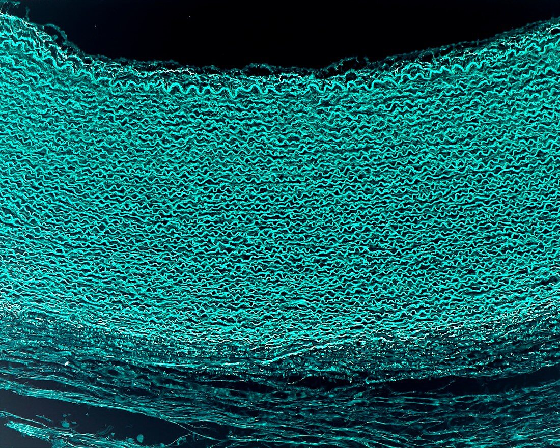
| Cross-section of a human aorta showing the layers of its wall, light micrograph. The tunica media, rich in elastic sheets or lamellae, occupies most of the wall. In the lower part of the image, the tunica adventitia is placed, looser in appearance and devoid of elastic lamellae. Orcein stain for elastin. The image is, in reality, the negative of the microscopic image. In this way, the elastic elements, which now appear in bright green, stand out even more clearly. Magnification: x90 when printed at 10 centimetres across. | |
| Lizenzart: | Lizenzpflichtig |
| Credit: | Science Photo Library / JOSE CALVO |
| Bildgröße: | 4674 px × 3739 px |
| Modell-Rechte: | nicht erforderlich |
| Eigentums-Rechte: | nicht erforderlich |
| Restrictions: | - |
Preise für dieses Bild ab 15 €
Universitäten & Organisationen
(Informationsmaterial Digital, Informationsmaterial Print, Lehrmaterial Digital etc.)
ab 15 €
Redaktionell
(Bücher, Bücher: Sach- und Fachliteratur, Digitale Medien (redaktionell) etc.)
ab 30 €
Werbung
(Anzeigen, Aussenwerbung, Digitale Medien, Fernsehwerbung, Karten, Werbemittel, Zeitschriften etc.)
ab 55 €
Handelsprodukte
(bedruckte Textilie, Kalender, Postkarte, Grußkarte, Verpackung etc.)
ab 75 €
Pauschalpreise
Rechtepakete für die unbeschränkte Bildnutzung in Print oder Online
ab 495 €
Keywords
- Adventitia,
- Anatomie,
- anatomisch,
- Arterie,
- arteriell,
- Arteriole,
- Ballaststoff,
- befleckt,
- Bindegewebe,
- Biologie,
- biologisch,
- Blut,
- Blutfluss,
- Blutgefäß,
- Blutgefäße,
- Blutzelle,
- Elastin,
- elastische Fasern,
- Fasern,
- Gefäß,
- Gefäße,
- gesund,
- Gesundheit,
- Gesundheitswesen,
- Glatt,
- Histologie,
- histologisch,
- Kreislauf,
- Lichtmikroskop,
- lichtmikroskopische Aufnahme,
- Medien,
- Mensch,
- menschlicher Körper,
- menschliches Gewebe,
- Muskulös,
- normal,
- rote Blutkörperchen,
- Schicht,
- Schichten,
- Sektion,
- Tunica intima,
- vaskulär,
- Vene