Compact bone, light micrograph
Bildnummer 12360846
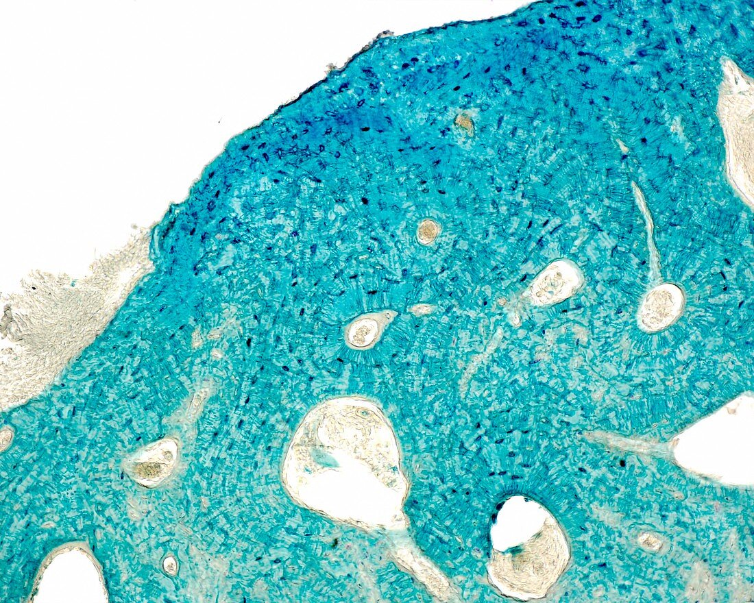
| Fragment of a compact bone stained with the Schmorl technique, light micrograph. With this technique, osteocytes and their processes are stained blue. Unstained spaces of different sizes are also observed. The smallest and more rounded are Haversian canals. They are surrounded by osteocytes arranged in concentric layers forming the osteons or Haversian systems. Larger spaces are reabsorption cavities. Magnification: x90 when printed at 10 centimetres across. | |
| Lizenzart: | Lizenzpflichtig |
| Credit: | Science Photo Library / JOSE CALVO |
| Bildgröße: | 4674 px × 3739 px |
| Modell-Rechte: | nicht erforderlich |
| Eigentums-Rechte: | nicht erforderlich |
| Restrictions: | - |
Preise für dieses Bild ab 15 €
Universitäten & Organisationen
(Informationsmaterial Digital, Informationsmaterial Print, Lehrmaterial Digital etc.)
ab 15 €
Redaktionell
(Bücher, Bücher: Sach- und Fachliteratur, Digitale Medien (redaktionell) etc.)
ab 30 €
Werbung
(Anzeigen, Aussenwerbung, Digitale Medien, Fernsehwerbung, Karten, Werbemittel, Zeitschriften etc.)
ab 55 €
Handelsprodukte
(bedruckte Textilie, Kalender, Postkarte, Grußkarte, Verpackung etc.)
ab 75 €
Pauschalpreise
Rechtepakete für die unbeschränkte Bildnutzung in Print oder Online
ab 495 €
Keywords
- Anatomie,
- anatomisch,
- befleckt,
- Biologie,
- biologisch,
- gesund,
- Gewebe,
- Histologie,
- histologisch,
- Kalzium,
- Knochen,
- Kollagen,
- Lichtmikroskop,
- lichtmikroskopische Aufnahme,
- Matrix,
- menschlicher Körper,
- Mikroskopie,
- Mineral,
- Osteon,
- Osteozyten,
- Sektion,
- sektioniert,
- Skelettknochen,
- Zelle,
- Zellen,
- Zytologie,
- Zytologisch