Pancreatic serous acini, light micrograph
Bildnummer 12360840
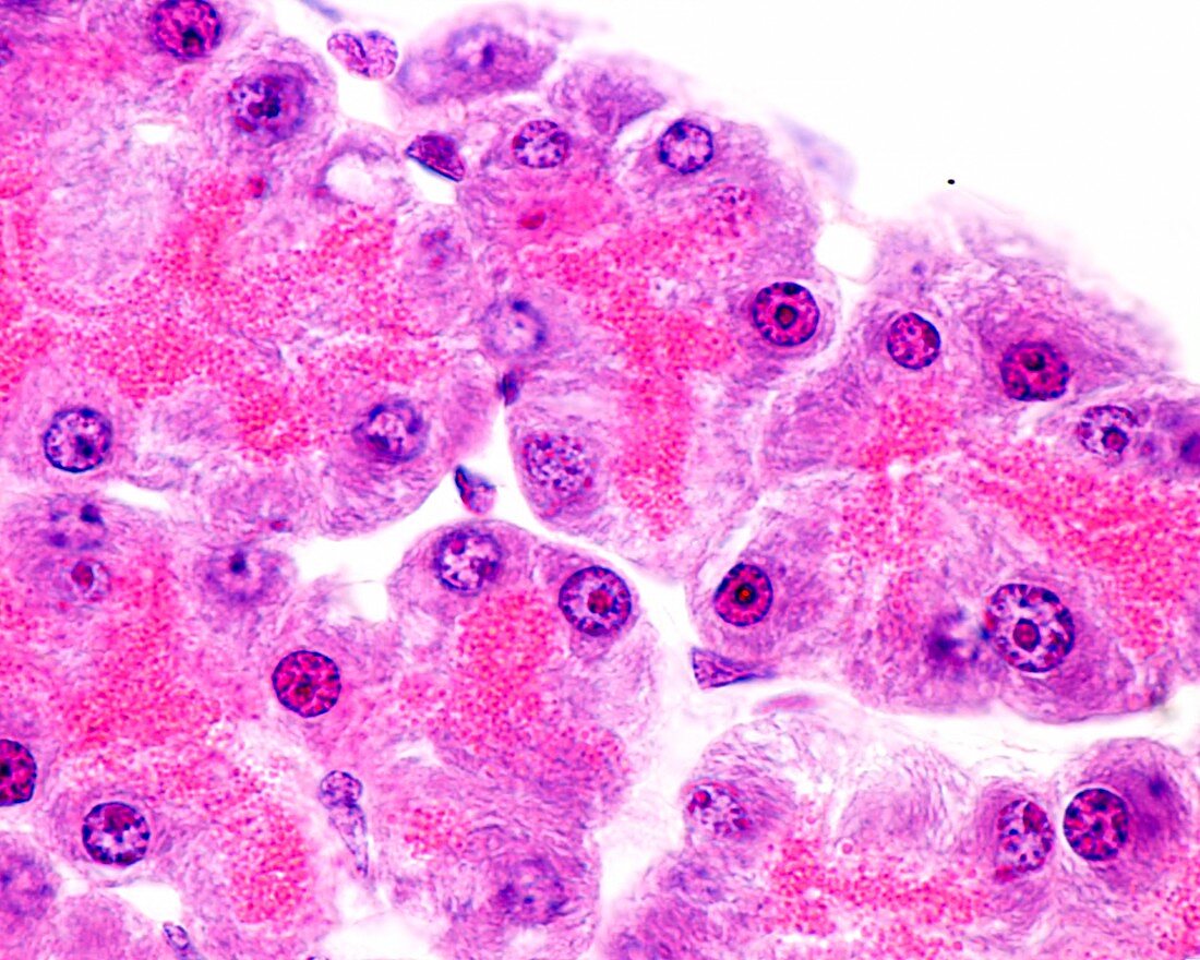
| High magnification light micrograph showing pancreatic serous acini. The two typical zones of a serous cell are clearly distinguished. At the base, the basophilic (dark purple) staining is due to the abundant rough endoplasmic reticulum present in this cell type. The nucleus has a very developed nucleolus. The apical cytoplasm is filled with serous granules, intensely stained by eosin (pink) due to its richness in proteins (enzymes). Note how the secretory units visible in the image are not spherical, but elongated and even branched (tubuloacinar gland). Magnification: x900 when printed at 10 centimetres across. | |
| Lizenzart: | Lizenzpflichtig |
| Credit: | Science Photo Library / JOSE CALVO |
| Bildgröße: | 4674 px × 3739 px |
| Modell-Rechte: | nicht erforderlich |
| Eigentums-Rechte: | nicht erforderlich |
| Restrictions: | - |
Preise für dieses Bild ab 15 €
Universitäten & Organisationen
(Informationsmaterial Digital, Informationsmaterial Print, Lehrmaterial Digital etc.)
ab 15 €
Redaktionell
(Bücher, Bücher: Sach- und Fachliteratur, Digitale Medien (redaktionell) etc.)
ab 30 €
Werbung
(Anzeigen, Aussenwerbung, Digitale Medien, Fernsehwerbung, Karten, Werbemittel, Zeitschriften etc.)
ab 55 €
Handelsprodukte
(bedruckte Textilie, Kalender, Postkarte, Grußkarte, Verpackung etc.)
ab 75 €
Pauschalpreise
Rechtepakete für die unbeschränkte Bildnutzung in Print oder Online
ab 495 €