Sympathetic neuron, TEM
Bildnummer 12360610
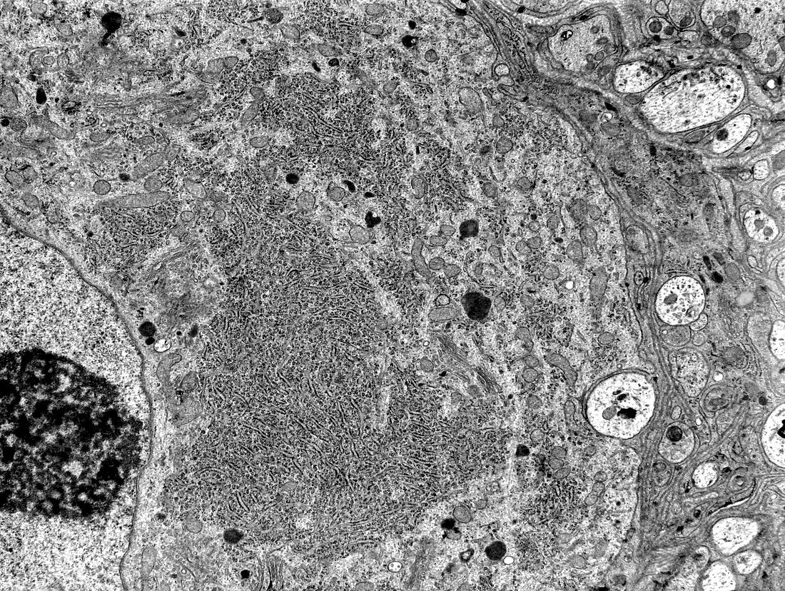
| Transmission electron micrograph (TEM) showing the cell body of a sympathetic nerve cell. The nucleus with a big nucleolus is at the left border. In the cytoplasm, a highly developed rough endoplasmic reticulum and some lysosomes stand out. Near the right border, glial cells and some nerve fibres can be seen. | |
| Lizenzart: | Lizenzpflichtig |
| Credit: | Science Photo Library / JOSE CALVO |
| Bildgröße: | 3549 px × 2670 px |
| Modell-Rechte: | nicht erforderlich |
| Eigentums-Rechte: | nicht erforderlich |
| Restrictions: | - |
Preise für dieses Bild ab 15 €
Universitäten & Organisationen
(Informationsmaterial Digital, Informationsmaterial Print, Lehrmaterial Digital etc.)
ab 15 €
Redaktionell
(Bücher, Bücher: Sach- und Fachliteratur, Digitale Medien (redaktionell) etc.)
ab 30 €
Werbung
(Anzeigen, Aussenwerbung, Digitale Medien, Fernsehwerbung, Karten, Werbemittel, Zeitschriften etc.)
ab 55 €
Handelsprodukte
(bedruckte Textilie, Kalender, Postkarte, Grußkarte, Verpackung etc.)
ab 75 €
Pauschalpreise
Rechtepakete für die unbeschränkte Bildnutzung in Print oder Online
ab 495 €
Keywords
- Atomhülle,
- Atomkern,
- Biologie,
- biologisch,
- Fehlfarbe,
- gefärbt,
- Gehirn,
- Gliazellen,
- Golgi-Apparat,
- Histologie,
- histologisch,
- Kerne,
- Mikrofotografie,
- Mikroskop,
- Mikroskopie,
- Mitochondrien,
- Mitochondrion,
- Nervenzelle,
- Neuron,
- tem,
- Transmissionselektronenmikroskop,
- transmissionselektronenmikroskopische Aufnahme,
- Ultrastruktur,
- Zelle,
- zentrales Nervensystem,
- Zytologie,
- Zytologisch,
- Zytoplasma