Sympathetic neuron, TEM
Bildnummer 12360609
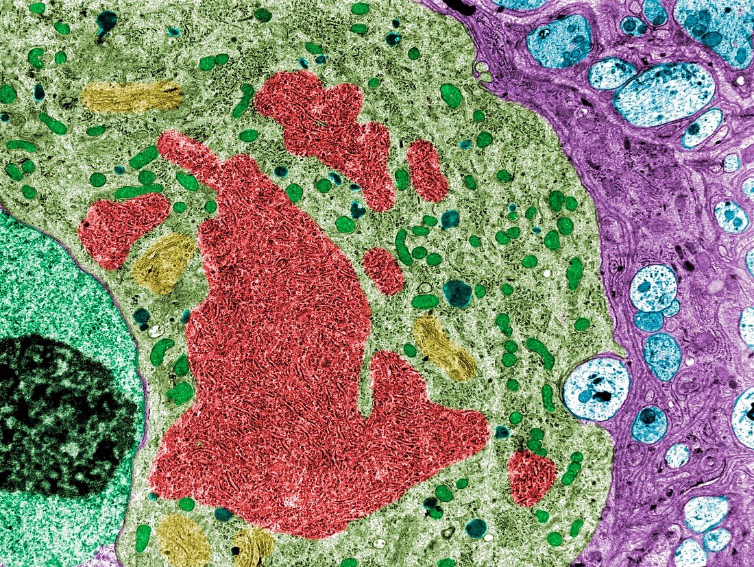
| False colour transmission electron micrograph (TEM) showing the cell body of a sympathetic nerve cell. The nucleus with a big nucleolus (light green) is at the left border. In the cytoplasm, a highly developed rough endoplasmic reticulum (brown). Golgi apparatus (yellow), mitochondria (dark green) and some lysosomes (dark blue) can also be seen. Near the right border, there are glial cells (pink) and some nerve fibres (light blue). | |
| Lizenzart: | Lizenzpflichtig |
| Credit: | Science Photo Library / JOSE CALVO |
| Bildgröße: | 3553 px × 2673 px |
| Modell-Rechte: | nicht erforderlich |
| Eigentums-Rechte: | nicht erforderlich |
| Restrictions: | - |
Preise für dieses Bild ab 15 €
Universitäten & Organisationen
(Informationsmaterial Digital, Informationsmaterial Print, Lehrmaterial Digital etc.)
ab 15 €
Redaktionell
(Bücher, Bücher: Sach- und Fachliteratur, Digitale Medien (redaktionell) etc.)
ab 30 €
Werbung
(Anzeigen, Aussenwerbung, Digitale Medien, Fernsehwerbung, Karten, Werbemittel, Zeitschriften etc.)
ab 55 €
Handelsprodukte
(bedruckte Textilie, Kalender, Postkarte, Grußkarte, Verpackung etc.)
ab 75 €
Pauschalpreise
Rechtepakete für die unbeschränkte Bildnutzung in Print oder Online
ab 495 €
Keywords
- Atomhülle,
- Atomkern,
- Biologie,
- biologisch,
- Fehlfarbe,
- gefärbt,
- Gehirn,
- Gliazellen,
- Golgi-Apparat,
- Histologie,
- histologisch,
- Kerne,
- Mikrofotografie,
- Mikroskop,
- Mikroskopie,
- Mitochondrien,
- Mitochondrion,
- Nervenzelle,
- Neuron,
- tem,
- Transmissionselektronenmikroskop,
- transmissionselektronenmikroskopische Aufnahme,
- Ultrastruktur,
- Zelle,
- zentrales Nervensystem,
- Zytologie,
- Zytologisch,
- Zytoplasma