Excised tumour in breast cancer surgery
Bildnummer 12347444
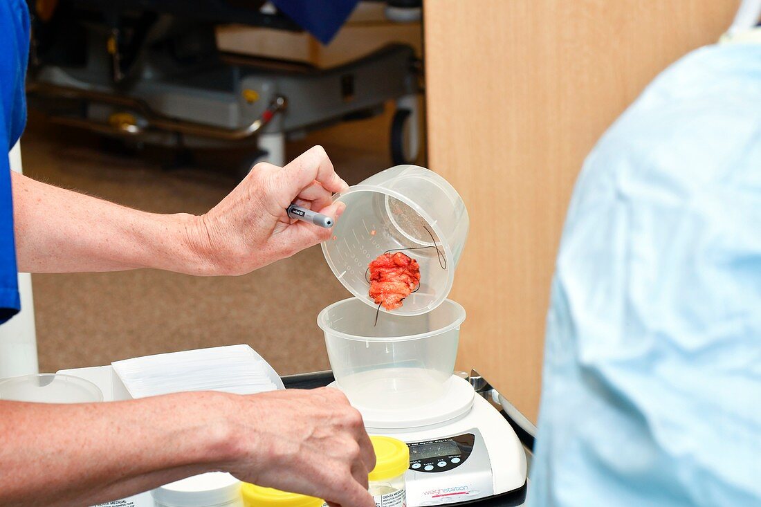
| Excised tumour in breast cancer surgery. Close-up of an excised and sutured tumour being weighed during breast cancer surgery on an 80-year-old woman. This operation involved the use of guide wires and radioactive tracers to accurately locate the malignant tissue to be removed. This surgery also involved operating on the cancer's sentinel lymph node. Following excision, the cancers are weighed. Sutures and staples inserted into the specimens help radiologists and histologists when examining them. Breast cancer is the most common cancer in women. The cancer can be effectively treated by surgery followed by a course of radiotherapy or anti-cancer drugs to destroy any remaining cancerous cells. | |
| Lizenzart: | Lizenzpflichtig |
| Credit: | Science Photo Library / Marazzi, Dr. P. |
| Bildgröße: | 5179 px × 3452 px |
| Modell-Rechte: | vorhanden |
| Eigentums-Rechte: | nicht erforderlich |
| Restrictions: | - |
Preise für dieses Bild ab 15 €
Universitäten & Organisationen
(Informationsmaterial Digital, Informationsmaterial Print, Lehrmaterial Digital etc.)
ab 15 €
Redaktionell
(Bücher, Bücher: Sach- und Fachliteratur, Digitale Medien (redaktionell) etc.)
ab 30 €
Werbung
(Anzeigen, Aussenwerbung, Digitale Medien, Fernsehwerbung, Karten, Werbemittel, Zeitschriften etc.)
ab 55 €
Handelsprodukte
(bedruckte Textilie, Kalender, Postkarte, Grußkarte, Verpackung etc.)
ab 75 €
Pauschalpreise
Rechtepakete für die unbeschränkte Bildnutzung in Print oder Online
ab 495 €
Keywords
- 2016,
- 21. Jahrhundert,
- abnormal,
- achtziger Jahre,
- Alt,
- Behandlung,
- Betrieb,
- britisch,
- Brustkrebs,
- Chirurg,
- Chirurgen,
- chirurgisch,
- Close-up,
- Container,
- Detail,
- eine Person,
- England,
- Englisch,
- Erwachsene,
- Europa,
- europäisch,
- Großbritannien,
- Hände,
- Handschuhe,
- Histopathologie,
- histopathologisch,
- klinisch,
- Kondition,
- Krankenhaus,
- Krankheit,
- Krebs,
- krebsartig,
- maligne,
- Malignom,
- Medizin,
- medizinisch,
- Menschen,
- Nähte,
- Onkologie,
- Operation,
- Operationssaal,
- Operationswunde,
- Person,
- Probe,
- Störung,
- Sutur,
- Tumor,
- ungesund,
- Vereinigtes Königreich,
- Waage,
- Wachstum,
- weiß,
- Wiegen