Trithigmostoma ciliate, LM
Bildnummer 12334234
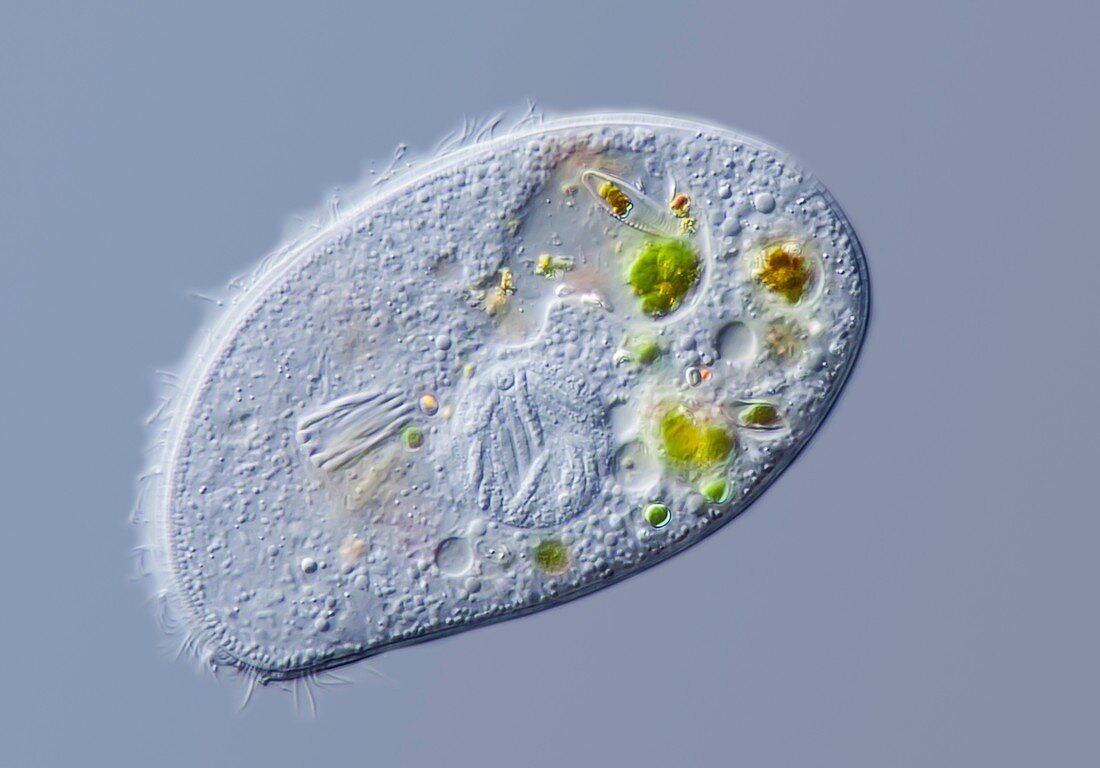
| Light micrograph of the cyrtophorid ciliate Trithigmostoma cucullus. Trithigmostoma feeds on Diatoms and other small algae, ingested via an oral basket, partially seen here on left side of the animal. Several green food vacuoles are visible at the right side of the animal. In the middle of the image the macronucleus is visible, in this case containing several parasitic bacteria. Microscopic contrast technique : Differential interference contrast. Magnification 650x when printed 10 centimetres wide. | |
| Lizenzart: | Lizenzpflichtig |
| Credit: | Science Photo Library / Guenther, Gerd |
| Bildgröße: | 5003 px × 3493 px |
| Modell-Rechte: | nicht erforderlich |
| Eigentums-Rechte: | nicht erforderlich |
| Restrictions: | - |
Preise für dieses Bild ab 15 €
Universitäten & Organisationen
(Informationsmaterial Digital, Informationsmaterial Print, Lehrmaterial Digital etc.)
ab 15 €
Redaktionell
(Bücher, Bücher: Sach- und Fachliteratur, Digitale Medien (redaktionell) etc.)
ab 30 €
Werbung
(Anzeigen, Aussenwerbung, Digitale Medien, Fernsehwerbung, Karten, Werbemittel, Zeitschriften etc.)
ab 55 €
Handelsprodukte
(bedruckte Textilie, Kalender, Postkarte, Grußkarte, Verpackung etc.)
ab 75 €
Pauschalpreise
Rechtepakete für die unbeschränkte Bildnutzung in Print oder Online
ab 495 €
Keywords
- Bakterien,
- Biologie,
- biologisch,
- Blauer Hintergrund,
- Ciliat,
- Ciliaten,
- DIC,
- Differenzialinterferenzkontrast,
- einzellig,
- Fauna,
- Lichtmikroskop,
- lichtmikroskopische Aufnahme,
- Mikrobiologie,
- mikrobiologisch,
- Mikroorganismen,
- Mikroorganismus,
- Mikroskop,
- Mikroskopie,
- mikroskopisch,
- Natur,
- Parasiten,
- Protist,
- Protisten,
- Protozoen,
- Protozoon,
- Single,
- Süßwasser,
- Tier,
- Tiere,
- Tierwelt,
- Wasser-,
- Wimpern,
- Zoologie,
- zoologisch