Neck bones and spinal cord, CT scan
Bildnummer 12303413
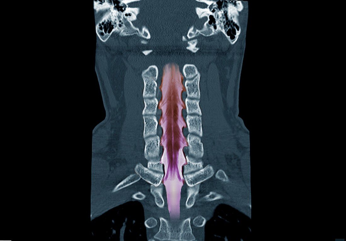
| Neck bones and spinal cord. Coloured coronal computed tomography (CT) scan of the neck bones and spinal cord of a 43-year-old man. This scan uses a contrast medium to highlight the spinal cord and the nerves branching off it, in a technique known as myelography. The scan shows the normal anatomy of the cervical (neck) region of the spinal cord, the spinal nerve roots, and the anatomy of the cervical vertebrae (spinal bones). | |
| Lizenzart: | Lizenzpflichtig |
| Credit: | Science Photo Library / Zephyr |
| Bildgröße: | 5001 px × 3495 px |
| Modell-Rechte: | nicht erforderlich |
| Eigentums-Rechte: | nicht erforderlich |
| Restrictions: | - |
Preise für dieses Bild ab 15 €
Universitäten & Organisationen
(Informationsmaterial Digital, Informationsmaterial Print, Lehrmaterial Digital etc.)
ab 15 €
Redaktionell
(Bücher, Bücher: Sach- und Fachliteratur, Digitale Medien (redaktionell) etc.)
ab 30 €
Werbung
(Anzeigen, Aussenwerbung, Digitale Medien, Fernsehwerbung, Karten, Werbemittel, Zeitschriften etc.)
ab 55 €
Handelsprodukte
(bedruckte Textilie, Kalender, Postkarte, Grußkarte, Verpackung etc.)
ab 75 €
Pauschalpreise
Rechtepakete für die unbeschränkte Bildnutzung in Print oder Online
ab 495 €
Keywords
- 40er Jahre,
- Anatomie,
- anatomisch,
- anterior,
- Biologie,
- biologisch,
- Computertomographie,
- CT-Scan,
- Erwachsene,
- farbig,
- Frontal,
- Gebärmutterhals-,
- gefärbt,
- gesund,
- Gewebe,
- Hals,
- Knochen,
- Kontrastmittel,
- Mann,
- Männlich,
- menschlicher Körper,
- Myelogramm,
- Myelographie,
- Nerv,
- Neurologie,
- neurologisch,
- Niemand,
- normal,
- Rücken,
- Rückenmark,
- Rückgrat,
- Scanner,
- schwarzer Hintergrund,
- Stirnbein,
- Vierziger Jahre,
- Wirbel,
- Wirbelsäule,
- Wirbelsäulen-,
- zentrales Nervensystem