Prostate cancer, abdominal CT scan
Bildnummer 12303352
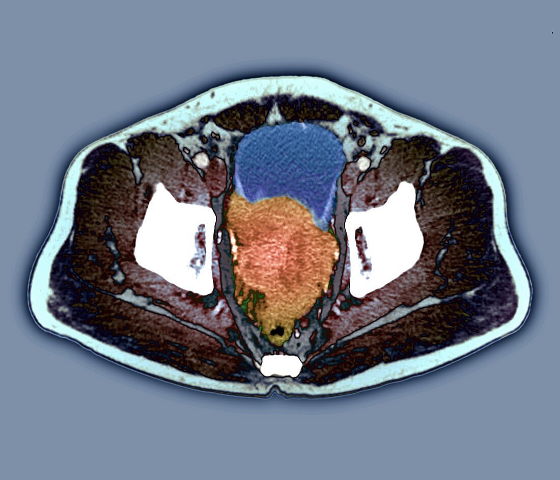
| Prostate cancer. Coloured axial computed tomography (CT) scan through the abdomen of a 88-year-old man with prostate cancer. The scan is centred on the bladder (blue), with the spine (white, lower centre) and hip bones (white, left and right) also visible. At centre is a large malignant tumour (orange) that has formed in the prostate gland and spread to the rectum and the bladder wall. | |
| Lizenzart: | Lizenzpflichtig |
| Credit: | Science Photo Library / Zephyr |
| Bildgröße: | 4518 px × 3868 px |
| Modell-Rechte: | nicht erforderlich |
| Eigentums-Rechte: | nicht erforderlich |
| Restrictions: | - |
Preise für dieses Bild ab 15 €
Universitäten & Organisationen
(Informationsmaterial Digital, Informationsmaterial Print, Lehrmaterial Digital etc.)
ab 15 €
Redaktionell
(Bücher, Bücher: Sach- und Fachliteratur, Digitale Medien (redaktionell) etc.)
ab 30 €
Werbung
(Anzeigen, Aussenwerbung, Digitale Medien, Fernsehwerbung, Karten, Werbemittel, Zeitschriften etc.)
ab 55 €
Handelsprodukte
(bedruckte Textilie, Kalender, Postkarte, Grußkarte, Verpackung etc.)
ab 75 €
Pauschalpreise
Rechtepakete für die unbeschränkte Bildnutzung in Print oder Online
ab 495 €
Keywords
- Abdomen,
- abnormal,
- achtziger Jahre,
- Alt,
- älter,
- ausgeschnitten,
- Ausschnitte,
- axial,
- Bauch,
- Blase,
- Computertomographie,
- CT-Scan,
- Drüse,
- Enddarm,
- Erwachsene,
- farbig,
- geduldig,
- gefärbt,
- Gewebe,
- grauer Hintergrund,
- Kondition,
- Krankheit,
- Krebs,
- krebsartig,
- maligne,
- Malignom,
- Mann,
- Männlich,
- Medizin,
- medizinisch,
- menschlicher Körper,
- Niemand,
- Onkologie,
- Organ,
- primär,
- Prostata,
- Prostatakrebs,
- Scanner,
- Sektion,
- sektioniert,
- sekundär,
- Störung,
- Tumor,
- ungesund,
- Verbreitung,
- Wachstum