Human brain anatomy, MRI scans
Bildnummer 12303340
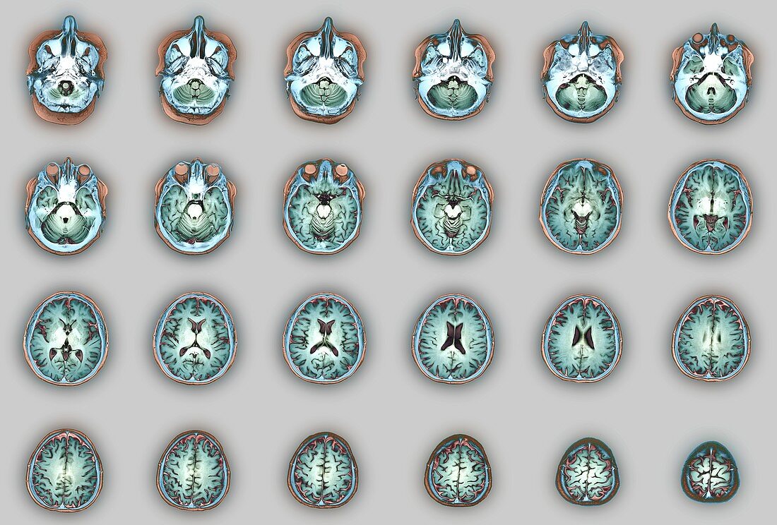
| Human brain anatomy. Series of coloured axial magnetic resonance imaging (MRI) scans of the brain of a 33-year-old man. The scans pass horizontally through the brain, with the sequence of 24 images running in rows from the base of the brain (top left) to the top of the brain (bottom right). Structures shown include the cerebellum (rear of brain in images 2-6), the nose (first 7 images), the eyes (images 5-8), and the ventricles of the brain (between the two hemispheres, images 13-16). These MRI scans are T2-weighted scans, with the use of gadolinium as a contrast medium. | |
| Lizenzart: | Lizenzpflichtig |
| Credit: | Science Photo Library / Zephyr |
| Bildgröße: | 5083 px × 3438 px |
| Modell-Rechte: | nicht erforderlich |
| Eigentums-Rechte: | nicht erforderlich |
| Restrictions: | - |
Preise für dieses Bild ab 15 €
Universitäten & Organisationen
(Informationsmaterial Digital, Informationsmaterial Print, Lehrmaterial Digital etc.)
ab 15 €
Redaktionell
(Bücher, Bücher: Sach- und Fachliteratur, Digitale Medien (redaktionell) etc.)
ab 30 €
Werbung
(Anzeigen, Aussenwerbung, Digitale Medien, Fernsehwerbung, Karten, Werbemittel, Zeitschriften etc.)
ab 55 €
Handelsprodukte
(bedruckte Textilie, Kalender, Postkarte, Grußkarte, Verpackung etc.)
ab 75 €
Pauschalpreise
Rechtepakete für die unbeschränkte Bildnutzung in Print oder Online
ab 495 €
Keywords
- 30er Jahre,
- Abschnitte,
- Anatomie,
- anatomisch,
- Augen,
- ausgeschnitten,
- Ausschnitte,
- axial,
- Base,
- Biologie,
- biologisch,
- dreißiger Jahre,
- Erwachsene,
- farbig,
- gefärbt,
- Gehirn,
- gesund,
- grauer Hintergrund,
- Kleinhirn,
- Kontrastmittel,
- Kopf,
- Magnetresonanztomografie,
- Mann,
- Männlich,
- medizinisch,
- menschlicher Körper,
- MRT-Untersuchung,
- Nase,
- Nerven,
- neural,
- Neurologie,
- neurologisch,
- Niemand,
- normal,
- Reihenfolge,
- Scanner,
- Scheiben,
- Sektion,
- sektioniert,
- Serie,
- untere