Eye anatomy and muscles, MRI scan
Bildnummer 12303326
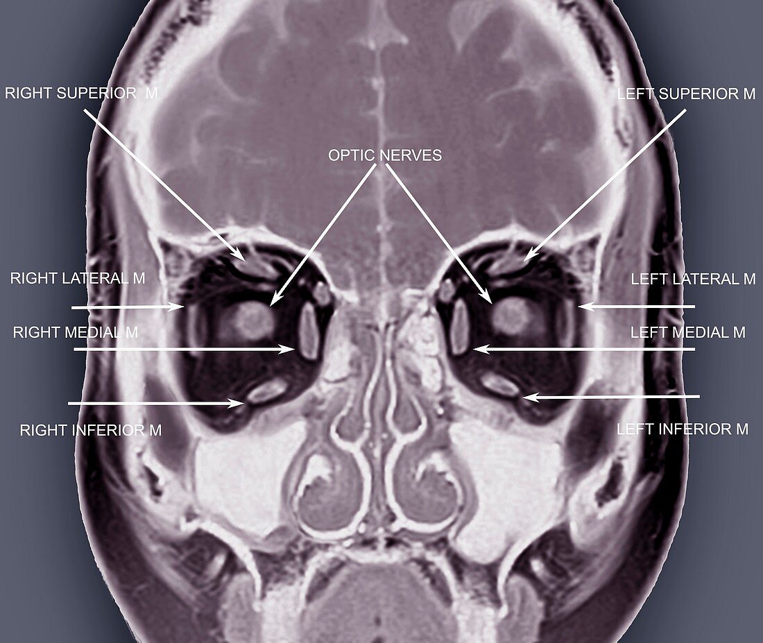
| Eye anatomy and muscles. Labelled coloured coronal magnetic resonance imaging (MRI) scan passing vertically through the head of a 33-year-old man, seen from the front, with the scan passing just behind the eyes. This scan shows some of the normal structures of the eyes and their support tissues, including the eye muscles (grey blocks) and the optic nerves. The section is at the level of a structure known as Tenon's capsule or the bulbar sheath. The muscles labelled here are the superior, lateral, medial and inferior orbital muscles of each eye. The optic nerves are also labelled. Parts of the brain and nasal sinuses are also visible on this scan. This is a T1-weighted scan, with injection of gadolinium contrast medium. For an unlabelled version of this scan, see image C033/7449. | |
| Lizenzart: | Lizenzpflichtig |
| Credit: | Science Photo Library / Zephyr |
| Bildgröße: | 4554 px × 3838 px |
| Modell-Rechte: | nicht erforderlich |
| Eigentums-Rechte: | nicht erforderlich |
| Restrictions: | - |
Preise für dieses Bild ab 15 €
Universitäten & Organisationen
(Informationsmaterial Digital, Informationsmaterial Print, Lehrmaterial Digital etc.)
ab 15 €
Redaktionell
(Bücher, Bücher: Sach- und Fachliteratur, Digitale Medien (redaktionell) etc.)
ab 30 €
Werbung
(Anzeigen, Aussenwerbung, Digitale Medien, Fernsehwerbung, Karten, Werbemittel, Zeitschriften etc.)
ab 55 €
Handelsprodukte
(bedruckte Textilie, Kalender, Postkarte, Grußkarte, Verpackung etc.)
ab 75 €
Pauschalpreise
Rechtepakete für die unbeschränkte Bildnutzung in Print oder Online
ab 495 €
Keywords
- 30er Jahre,
- Anatomie,
- anatomisch,
- Auge,
- Augen,
- Augen-,
- Augenheilkunde,
- beschriftet,
- Biologie,
- biologisch,
- dreißiger Jahre,
- Erwachsene,
- Etikette,
- Etiketten,
- farbig,
- gefärbt,
- Gehirn,
- Gesicht,
- gesund,
- Gewebe,
- Kontrastmittel,
- Kopf,
- links,
- Magnetresonanztomografie,
- Mann,
- Männlich,
- menschlicher Körper,
- MRT-Untersuchung,
- Muskel,
- Muskeln,
- Nasal-,
- Nebenhöhlen,
- Niemand,
- normal,
- okular,
- Orbital,
- Organ,
- Recht,
- Scanner,
- schwarzer Hintergrund,
- seitlich,
- Sektion,
- sektioniert,
- Sinus,
- Stirnbein,
- Text,
- überlegen,
- Umlaufbahnen