Implantation of stent in aorta, X-ray
Bildnummer 12302930
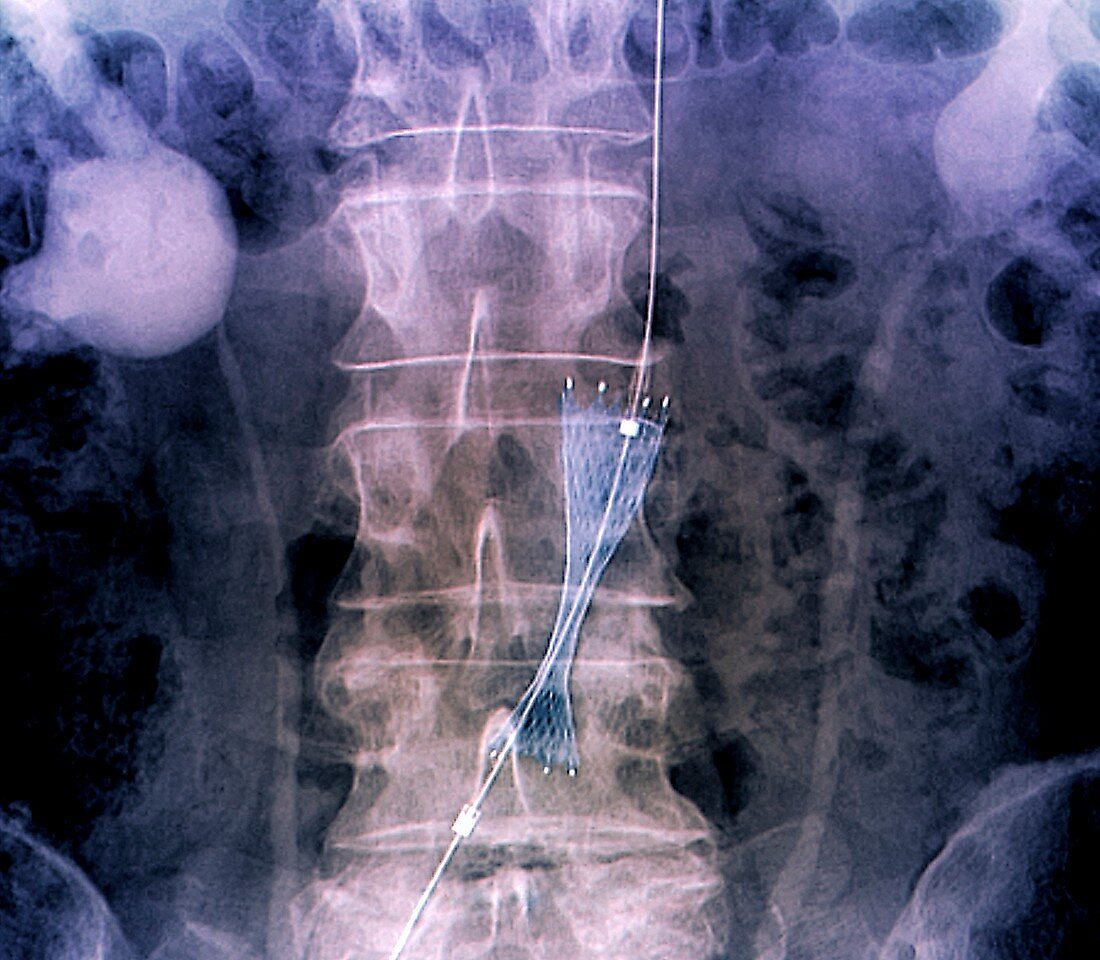
| Implantation of stent in aorta. Coloured frontal X-ray of a stent (mesh at lower centre) being implanted by an angioplasty catheter in the sub-renal section of the aorta of a 67-year-old man. The stent is being implanted to treat atheromatous lesions in the aorta that have led to a form of aortic dissection (separation of the layers of the aorta wall). The stent is a self-expansive endoprosthesis, which will be shaped in position using expansion balloons. The text label identifies the stent as a sinus-XL type, 20-60 millimetres in size. The spine is seen down centre, and the kidneys are at upper left and upper right. For the implantation of this stent, see images C033/6655 to C033/6657. | |
| Lizenzart: | Lizenzpflichtig |
| Credit: | Science Photo Library / Zephyr |
| Bildgröße: | 4474 px × 3906 px |
| Modell-Rechte: | nicht erforderlich |
| Eigentums-Rechte: | nicht erforderlich |
| Restrictions: | - |
Preise für dieses Bild ab 15 €
Universitäten & Organisationen
(Informationsmaterial Digital, Informationsmaterial Print, Lehrmaterial Digital etc.)
ab 15 €
Redaktionell
(Bücher, Bücher: Sach- und Fachliteratur, Digitale Medien (redaktionell) etc.)
ab 30 €
Werbung
(Anzeigen, Aussenwerbung, Digitale Medien, Fernsehwerbung, Karten, Werbemittel, Zeitschriften etc.)
ab 55 €
Handelsprodukte
(bedruckte Textilie, Kalender, Postkarte, Grußkarte, Verpackung etc.)
ab 75 €
Pauschalpreise
Rechtepakete für die unbeschränkte Bildnutzung in Print oder Online
ab 495 €
Keywords
- 60er Jahre,
- Abdomen,
- Angioplastie,
- Aorta,
- Arterie,
- Atherosklerose,
- Behandlung,
- beschriftet,
- chirurgisch,
- Erwachsene,
- Etikette,
- farbig,
- geduldig,
- gefärbt,
- Implantat,
- Kreislauf,
- Mann,
- Männlich,
- Medizin,
- medizinisch,
- menschlicher Körper,
- Niemand,
- Nieren-,
- Operation,
- Prothese,
- Radiographie,
- Röntgen,
- Röntgengerät,
- sechziger Jahre,
- Senior,
- Stent,
- thorakal,
- Thorax,
- vaskulär