Final position of stent in aorta, X-ray
Bildnummer 12302496
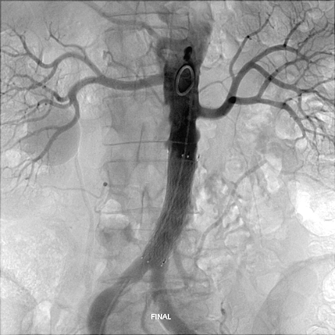
| Final position of stent in aorta. Frontal X-ray of a stent (mesh at lower centre) after being positioned and expanded by angioplasty catheters and balloons inside the sub-renal section of the aorta of a 67-year-old man. The stent was implanted to treat atheromatous lesions in the aorta that have led to a form of aortic dissection (separation of the layers of the aorta wall). The stent is a self-expansive endoprosthesis, which was shaped in position using expansion balloons. The stent is correctly placed and expanded to support the aorta wall. The spine is seen down centre, and the kidneys (and the renal blood vessels) are at upper left and upper right. For the implantation of this stent, see images C033/6619 to C033/6621. | |
| Lizenzart: | Lizenzpflichtig |
| Credit: | Science Photo Library / Zephyr |
| Bildgröße: | 4180 px × 4180 px |
| Modell-Rechte: | nicht erforderlich |
| Eigentums-Rechte: | nicht erforderlich |
| Restrictions: | - |
Preise für dieses Bild ab 15 €
Universitäten & Organisationen
(Informationsmaterial Digital, Informationsmaterial Print, Lehrmaterial Digital etc.)
ab 15 €
Redaktionell
(Bücher, Bücher: Sach- und Fachliteratur, Digitale Medien (redaktionell) etc.)
ab 30 €
Werbung
(Anzeigen, Aussenwerbung, Digitale Medien, Fernsehwerbung, Karten, Werbemittel, Zeitschriften etc.)
ab 55 €
Handelsprodukte
(bedruckte Textilie, Kalender, Postkarte, Grußkarte, Verpackung etc.)
ab 75 €
Pauschalpreise
Rechtepakete für die unbeschränkte Bildnutzung in Print oder Online
ab 495 €
Keywords
- 60er Jahre,
- Abdomen,
- Angioplastie,
- Aorta,
- Arterie,
- Atherosklerose,
- Behandlung,
- beschriftet,
- chirurgisch,
- Einfarbig,
- Erwachsene,
- Etikette,
- geduldig,
- Implantat,
- Kreislauf,
- Mann,
- Männlich,
- Medizin,
- medizinisch,
- menschlicher Körper,
- Niemand,
- Nieren-,
- Operation,
- Prothese,
- Radiographie,
- Röntgen,
- Röntgengerät,
- Schwarz und weiß,
- sechziger Jahre,
- Senior,
- Stent,
- thorakal,
- Thorax,
- vaskulär