Frog tympanum cell surface, SEM
Bildnummer 12301772
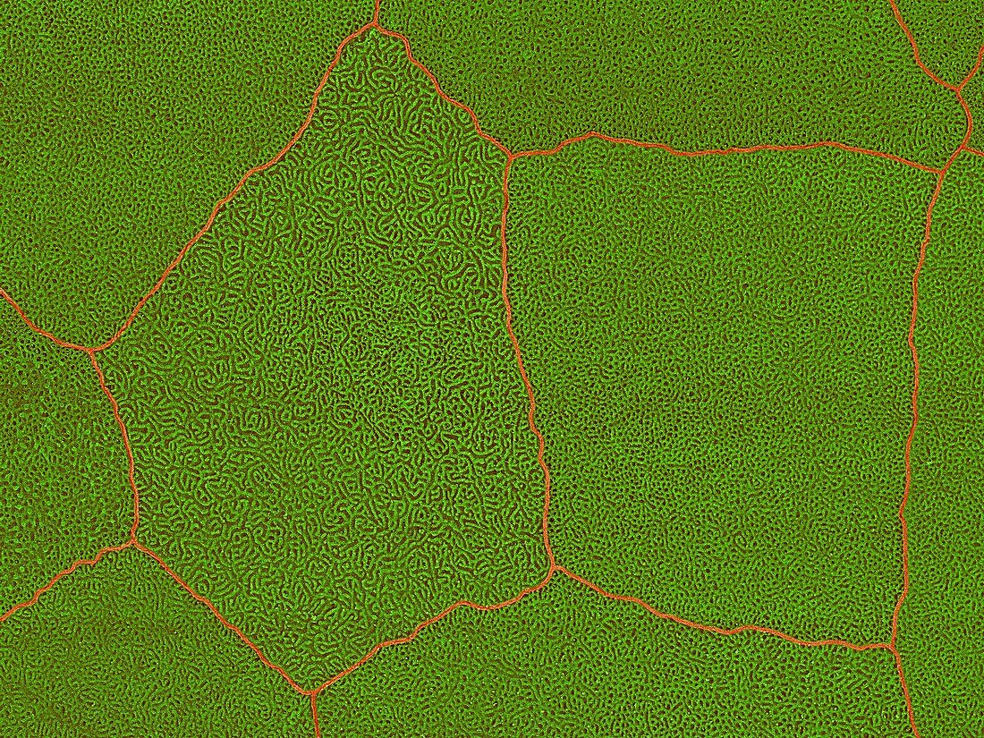
| Coloured scanning electron micrograph (SEM) Frog tympanic membrane cell surface (Eleutherodactylus coqui). Shown here is the surface of the frog tympanic membrane (ear membrane). Two individual epithelial cells with highly convoluted surface ridges are visible. In frogs a distinct tympanic fold wraps around the tympanum. The tympanum is composed of nonglandular skin cells (epithelial cells) and unlike the human eardrum it is situated externally on the frog (in most species behind the eye). It functions in a similar fashion to the human eardrum. There is a bone attached to the tympanic membrane that also attaches to the oval window of the inner ear. When sound waves strike the membrane vibrations are transferred via this bone to the fluid of the inner ear. This in turn causes receptors within the inner ear to be stimulated, sending a transmission to the brain for processing. Magnification: x440 when shortest axis printed at 25 | |
| Lizenzart: | Lizenzpflichtig |
| Credit: | Science Photo Library / DENNIS KUNKEL MICROSCOPY |
| Bildgröße: | 3413 px × 2560 px |
| Modell-Rechte: | nicht erforderlich |
| Eigentums-Rechte: | nicht erforderlich |
| Restrictions: | - |
Preise für dieses Bild ab 15 €
Universitäten & Organisationen
(Informationsmaterial Digital, Informationsmaterial Print, Lehrmaterial Digital etc.)
ab 15 €
Redaktionell
(Bücher, Bücher: Sach- und Fachliteratur, Digitale Medien (redaktionell) etc.)
ab 30 €
Werbung
(Anzeigen, Aussenwerbung, Digitale Medien, Fernsehwerbung, Karten, Werbemittel, Zeitschriften etc.)
ab 55 €
Handelsprodukte
(bedruckte Textilie, Kalender, Postkarte, Grußkarte, Verpackung etc.)
ab 75 €
Pauschalpreise
Rechtepakete für die unbeschränkte Bildnutzung in Print oder Online
ab 495 €
Keywords
- Amphibie,
- Baum,
- Chordatier,
- Elektron,
- Epithel,
- epithelial,
- Epithelien,
- Falten,
- farbig,
- Frosch,
- gefärbt,
- Haut,
- Hawaii,
- hawaiisch,
- Hören,
- Landwirtschaft,
- landwirtschaftlich,
- Membran,
- Mikrofotografie,
- Oberfläche,
- Ohr,
- Organ,
- Pest,
- REM,
- Scannen,
- Sinn,
- Spezies,
- Vibration,
- Vibrationen,
- Zelle,
- Zellen