Soil fungus Exophiala spinifera, SEM
Bildnummer 12300115
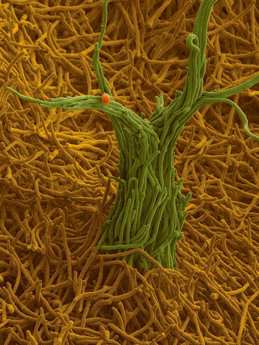
| Coloured scanning electron micrograph (SEM) of Exophiala spinifera is a dermatiaceous fungus that is widely distributed in soil, plants, water and decaying wood material. E. spinifera initially grows as yeast-like cells that are brownish to greenish-black in colour. The yeast colonies then develop compact hyphae which form a velvety mycelium. The mycelium develops short tufts of aerial hyphae (shown here). As well as being a common saprophyte in nature, Exophiala spinifera can cause various human subcutaneous infections including: mycetoma, cutaneous phaeohyphomycosis and chromoblastomycosis. These infections are often associated with organ transplants. Magnification: x400 when shortest axis printed at 25 millimetres. | |
| Lizenzart: | Lizenzpflichtig |
| Credit: | Science Photo Library / DENNIS KUNKEL MICROSCOPY |
| Bildgröße: | 2613 px × 3483 px |
| Modell-Rechte: | nicht erforderlich |
| Eigentums-Rechte: | nicht erforderlich |
| Restrictions: | - |
Preise für dieses Bild ab 15 €
Universitäten & Organisationen
(Informationsmaterial Digital, Informationsmaterial Print, Lehrmaterial Digital etc.)
ab 15 €
Redaktionell
(Bücher, Bücher: Sach- und Fachliteratur, Digitale Medien (redaktionell) etc.)
ab 30 €
Werbung
(Anzeigen, Aussenwerbung, Digitale Medien, Fernsehwerbung, Karten, Werbemittel, Zeitschriften etc.)
ab 55 €
Handelsprodukte
(bedruckte Textilie, Kalender, Postkarte, Grußkarte, Verpackung etc.)
ab 75 €
Pauschalpreise
Rechtepakete für die unbeschränkte Bildnutzung in Print oder Online
ab 495 €
Keywords
- Ascomycota,
- Ascosporen,
- asexuell,
- Boden,
- Elektron,
- farbig,
- Formen,
- Gastgeber,
- geduldig,
- gefärbt,
- Haut,
- Hyphe,
- Infektion,
- Konidien,
- Konidiophor,
- Körper,
- Krankheit,
- Krankheiten,
- Läsionen,
- Mensch,
- Mikrofotografie,
- Myzel,
- Organ,
- Pflanze,
- Pilz,
- Pilz-,
- Pilze,
- REM,
- Reproduktion,
- Saprophyt,
- Scannen,
- Schleim,
- sich durch Sporen vermehren,
- Sporen,
- Struktur,
- Strukturen,
- Transplantation,
- Wunde,
- Zelle,
- Zellen