Plasmodium falciparum, TEM
Bildnummer 12298356
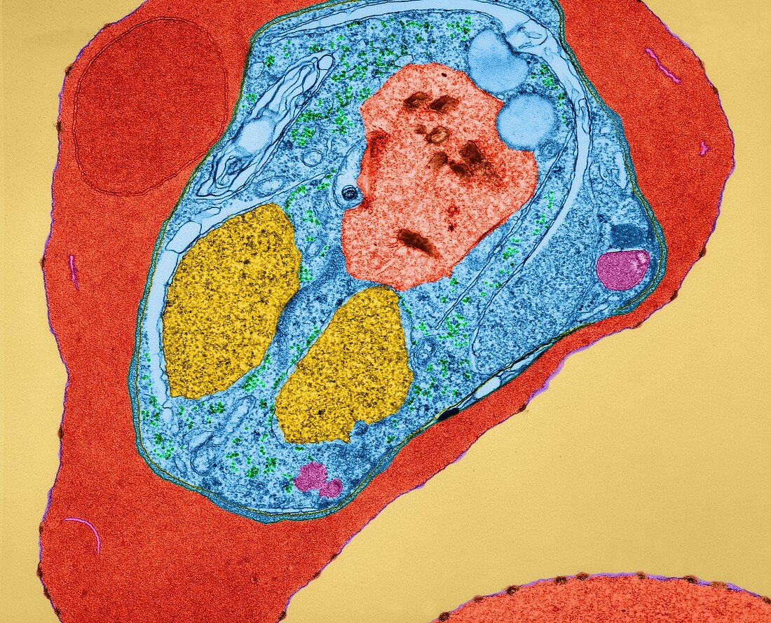
| Plasmodium falciparum plasmodial young schizont infecting an erythrocyte (red blood cell), coloured transmission electron micrograph (TEM). The red blood cell membrane has small distinct knobs (orange) that are characteristic of the infection by certain strains of Plasmodium falciparum. The young schizont stage cytoplasm has a distinct food vacuole (light orange) with hemozoin pigment granules (rust brown). The cytoplasm contains two nuclei (yellow), mitochondria (pink) and ribosomes (green). Maurer's clefts (light blue) can be seen in the haemoglobin-containing cytoplasm. Malaria is caused by Plasmodium spp., protozoa. It is spread to humans by Anopheles sp. mosquitoes. The plasmodial parasite reproduces asexually in red blood cells significantly destroying many of them. Release of mature Plasmodium merozoites results in further infection and produces bouts of shivering fever (paroxysms) and sweating that may be fatal. Magnification: x3, 810 when | |
| Lizenzart: | Lizenzpflichtig |
| Credit: | Science Photo Library / DENNIS KUNKEL MICROSCOPY |
| Bildgröße: | 5445 px × 4400 px |
| Modell-Rechte: | nicht erforderlich |
| Eigentums-Rechte: | nicht erforderlich |
| Restrictions: | - |
Preise für dieses Bild ab 15 €
Universitäten & Organisationen
(Informationsmaterial Digital, Informationsmaterial Print, Lehrmaterial Digital etc.)
ab 15 €
Redaktionell
(Bücher, Bücher: Sach- und Fachliteratur, Digitale Medien (redaktionell) etc.)
ab 30 €
Werbung
(Anzeigen, Aussenwerbung, Digitale Medien, Fernsehwerbung, Karten, Werbemittel, Zeitschriften etc.)
ab 55 €
Handelsprodukte
(bedruckte Textilie, Kalender, Postkarte, Grußkarte, Verpackung etc.)
ab 75 €
Pauschalpreise
Rechtepakete für die unbeschränkte Bildnutzung in Print oder Online
ab 495 €
Keywords
- asexuell,
- Blut,
- Bühne,
- chromalveolata,
- Elektron,
- Erythrozyt,
- Farbig,
- gefärbt,
- Infektion,
- infiziert,
- Krankheit,
- Leber,
- Mensch,
- Mikrofotografie,
- Parasit,
- parasitär,
- Plasmodium,
- Protozoen,
- Protozoon,
- rbc,
- Reproduktion,
- rot,
- Teilen,
- tem,
- Übertragung,
- Zelle,
- Zellen