Middle meningeal artery and haematoma, illustration
Bildnummer 12287356
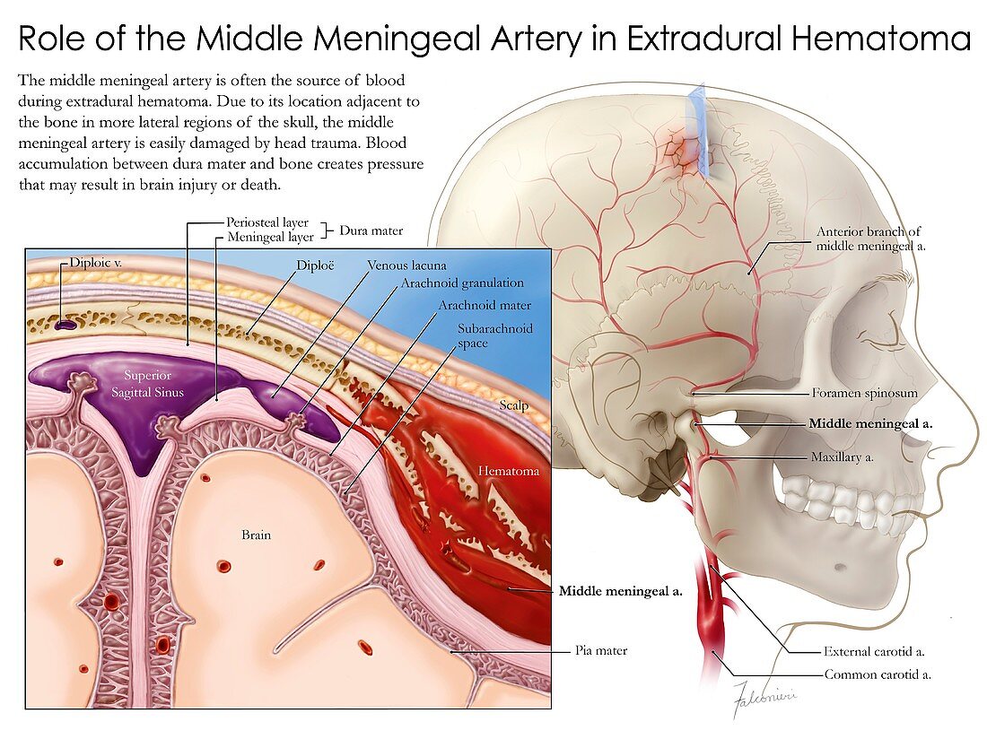
| Illustration of the anatomical relationship of the middle meningeal artery to the skull and meninges (brain membranes) and its role in extradural haematoma. The middle meningeal artery branches from the maxillary artery and serves the dura mater (outermost meninges) and the skullcap. The middle meningeal artery is covered by the weakest area of the skull and so is often damaged when the head receives a traumatic blow. Bleeding of the middle meningeal artery causes blood to accumulate between the skull and dura mater, which is known as an extradural haematoma. The haematoma increases pressure within the skull, compressing the brain and causing headache, drowsiness, vomiting, paralysis and in some cases death. | |
| Lizenzart: | Lizenzpflichtig |
| Credit: | Science Photo Library / VERONICA FALCONIERI HAYS |
| Bildgröße: | 5079 px × 3806 px |
| Modell-Rechte: | nicht erforderlich |
| Eigentums-Rechte: | nicht erforderlich |
| Restrictions: | - |
Preise für dieses Bild ab 15 €
Universitäten & Organisationen
(Informationsmaterial Digital, Informationsmaterial Print, Lehrmaterial Digital etc.)
ab 15 €
Redaktionell
(Bücher, Bücher: Sach- und Fachliteratur, Digitale Medien (redaktionell) etc.)
ab 30 €
Werbung
(Anzeigen, Aussenwerbung, Digitale Medien, Fernsehwerbung, Karten, Werbemittel, Zeitschriften etc.)
ab 55 €
Handelsprodukte
(bedruckte Textilie, Kalender, Postkarte, Grußkarte, Verpackung etc.)
ab 75 €
Pauschalpreise
Rechtepakete für die unbeschränkte Bildnutzung in Print oder Online
ab 495 €