False-colour SEM of filiform papillae and bacteria
Bildnummer 12261721
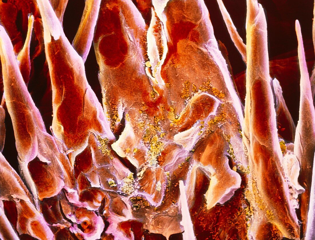
| False-colour scanning electron micrograph (SEM) of filiform papillae on the surface of the tongue. Filiform papillae, also known as conical papillae, are covered by stratified squamous epithelial cells apart from their tips which are formed by the fibrous protein keratin in order to increase the papillae's strength. Filiform papillae have mechanical and tactile functions. They form a rough surface which helps the mastication process. Each papilla contains nerve endings which transmit tactile information to the brain. At centre green coloured bacteria are also visible. Magnification: x925 at 6x7cm size. Magnification: x1360 at 4x5 inch size size. | |
| Lizenzart: | Lizenzpflichtig |
| Credit: | Science Photo Library / UNIVERSITY LA SAPIENZA"", ROME / DEPT. OF ANATOMY / PROF. P. MOTTA |
| Bildgröße: | 5262 px × 4004 px |
| Modell-Rechte: | nicht erforderlich |
| Eigentums-Rechte: | nicht erforderlich |
| Restrictions: | - |
Preise für dieses Bild ab 15 €
Universitäten & Organisationen
(Informationsmaterial Digital, Informationsmaterial Print, Lehrmaterial Digital etc.)
ab 15 €
Redaktionell
(Bücher, Bücher: Sach- und Fachliteratur, Digitale Medien (redaktionell) etc.)
ab 30 €
Werbung
(Anzeigen, Aussenwerbung, Digitale Medien, Fernsehwerbung, Karten, Werbemittel, Zeitschriften etc.)
ab 55 €
Handelsprodukte
(bedruckte Textilie, Kalender, Postkarte, Grußkarte, Verpackung etc.)
ab 75 €
Pauschalpreise
Rechtepakete für die unbeschränkte Bildnutzung in Print oder Online
ab 495 €