Skin anatomy, fluorescence light micrograph
Bildnummer 12249345
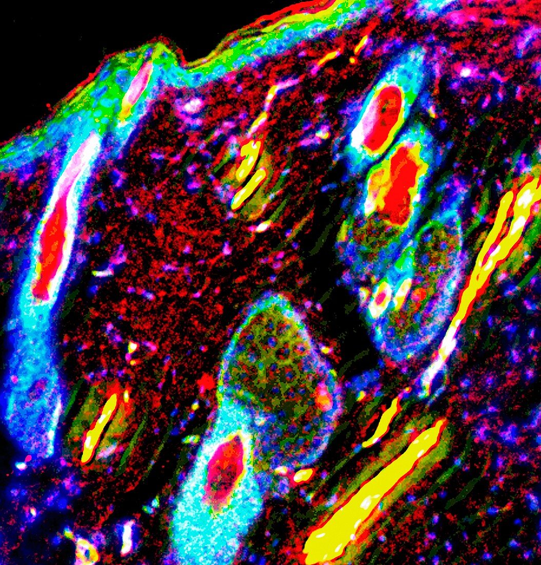
| Skin anatomy, fluorescence light micrograph. This image shows sebaceous glands (green), arrector pili muscles (yellow), hair follicles (white-blue), hair shafts (red), and the epidermis layer (top) with the stratum corneum (red) on top. Fluorescent markers have been used to highlight proteins in the cells. | |
| Lizenzart: | Lizenzpflichtig |
| Credit: | Science Photo Library / R. BICK, B. POINDEXTER, UT MEDICAL SCHOOL |
| Bildgröße: | 2895 px × 3020 px |
| Modell-Rechte: | nicht erforderlich |
| Eigentums-Rechte: | nicht erforderlich |
| Restrictions: | - |
Preise für dieses Bild ab 15 €
Universitäten & Organisationen
(Informationsmaterial Digital, Informationsmaterial Print, Lehrmaterial Digital etc.)
ab 15 €
Redaktionell
(Bücher, Bücher: Sach- und Fachliteratur, Digitale Medien (redaktionell) etc.)
ab 30 €
Werbung
(Anzeigen, Aussenwerbung, Digitale Medien, Fernsehwerbung, Karten, Werbemittel, Zeitschriften etc.)
ab 55 €
Handelsprodukte
(bedruckte Textilie, Kalender, Postkarte, Grußkarte, Verpackung etc.)
ab 75 €
Pauschalpreise
Rechtepakete für die unbeschränkte Bildnutzung in Print oder Online
ab 495 €
Keywords
- Anatomie,
- anatomisch,
- biologisch,
- dermal,
- Dermatologie,
- dermatologisch,
- Eiweiß,
- Epidermis,
- Fluoreszenz,
- fluoreszierend,
- gesund,
- Gewebe,
- Haut,
- Lichtmikroskop,
- lichtmikroskopische Aufnahme,
- menschlicher Körper,
- Niemand,
- normal,
- Proteine,
- Stratum corneum,
- Zellbilogie,
- Zelle,
- Zellen,
- Zytologie,
- Zytologisch