Fluorescence mapping of an oncogene
Bildnummer 12248853
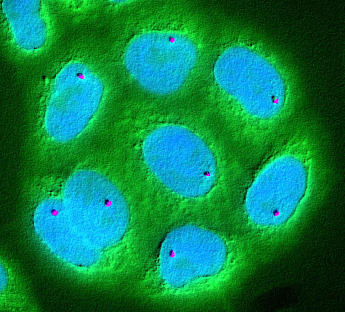
| Fluorescence mapping of an oncogene. Fluorescence light micrograph showing a close-up of cell nuclei, mapping the position of genes. This research aids understanding of what governs the genome. Here, a single gene called PEM (polymorphic epithelial mucin, purple) has been localized using fluorescence in-situ hybridization. DNA is stained blue, while the cell cytoplasm is stained green. Overexpression of PEM (also called MUC1) is often associated with colon, breast, ovarian, lung and pancreatic cancers. An oncogene is a gene that causes cancer. | |
| Lizenzart: | Lizenzpflichtig |
| Credit: | Science Photo Library / NATIONAL CANCER INSTITUTE / NCI Center for Cancer Research |
| Bildgröße: | 3117 px × 2824 px |
| Modell-Rechte: | nicht erforderlich |
| Eigentums-Rechte: | nicht erforderlich |
| Restrictions: |
|
Preise für dieses Bild ab 15 €
Universitäten & Organisationen
(Informationsmaterial Digital, Informationsmaterial Print, Lehrmaterial Digital etc.)
ab 15 €
Redaktionell
(Bücher, Bücher: Sach- und Fachliteratur, Digitale Medien (redaktionell) etc.)
ab 30 €
Werbung
(Anzeigen, Aussenwerbung, Digitale Medien, Fernsehwerbung, Karten, Werbemittel, Zeitschriften etc.)
ab 55 €
Handelsprodukte
(bedruckte Textilie, Kalender, Postkarte, Grußkarte, Verpackung etc.)
ab 75 €
Pauschalpreise
Rechtepakete für die unbeschränkte Bildnutzung in Print oder Online
ab 495 €
Keywords
- abnormal,
- Atomkern,
- Fluoreszenz,
- fluoreszierend,
- Forschung,
- Gen,
- Genetik,
- genetisch,
- Gewebe,
- Kondition,
- Krankheit,
- Krebs,
- krebsartig,
- Krebszelle,
- Lichtmikroskop,
- lichtmikroskopische Aufnahme,
- maligne,
- Malignom,
- Medizin,
- medizinisch,
- Niemand,
- Onkogen,
- Onkologie,
- pem,
- Störung,
- Tumor,
- ungesund,
- Zelle,
- Zellen,
- zellular