Ovarian cancer, optical tissue clearing
Bildnummer 12248830
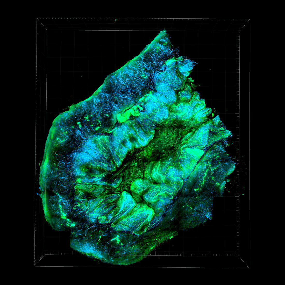
| Ovarian cancer. Optical tissue clearing image of tissue from an ovarian cancer, showing the interplay of collagen and blood vessels. This is done using second harmonic signals (blue) and autofluorescent signals (green; green fluorescent protein, GFP). This is a murine SKOV cell line tumour seeded with CD63+ cells, revealing the tumour-stromal interfaces that comprises the tumour microenvironment (TME). The image demonstrates the interplay of collagen II fibrils and blood vessels generated from angiogenesis. The tumour microenvironment is being studued because it plays a crucial role in helping cancers to grow and evade destruction. This image is of a mouse model of ovarian cancer. | |
| Lizenzart: | Lizenzpflichtig |
| Credit: | Science Photo Library / NATIONAL CANCER INSTITUTE / Comprehensive Cancer Center of Wake Forest University |
| Bildgröße: | 4228 px × 4228 px |
| Modell-Rechte: | nicht erforderlich |
| Eigentums-Rechte: | nicht erforderlich |
| Restrictions: |
|
Preise für dieses Bild ab 15 €
Universitäten & Organisationen
(Informationsmaterial Digital, Informationsmaterial Print, Lehrmaterial Digital etc.)
ab 15 €
Redaktionell
(Bücher, Bücher: Sach- und Fachliteratur, Digitale Medien (redaktionell) etc.)
ab 30 €
Werbung
(Anzeigen, Aussenwerbung, Digitale Medien, Fernsehwerbung, Karten, Werbemittel, Zeitschriften etc.)
ab 55 €
Handelsprodukte
(bedruckte Textilie, Kalender, Postkarte, Grußkarte, Verpackung etc.)
ab 75 €
Pauschalpreise
Rechtepakete für die unbeschränkte Bildnutzung in Print oder Online
ab 495 €
Keywords
- 3 dimensional,
- 3-dimensional,
- 3D,
- abnormal,
- Angiogenese,
- Bindegewebe,
- Blutgefäße,
- Dreidimensional,
- Eierstock,
- Eierstöcke,
- Forschung,
- Gewebe,
- GFP,
- Grün fluoreszierendes Protein,
- Kollagen,
- Kondition,
- Krankheit,
- Krebs,
- krebsartig,
- maligne,
- Malignom,
- Medizin,
- medizinisch,
- Niemand,
- Onkologie,
- Scanner,
- schwarzer Hintergrund,
- Störung,
- Tumor,
- ungesund,
- Wachstum