Breast cancer immune response
Bildnummer 12248822
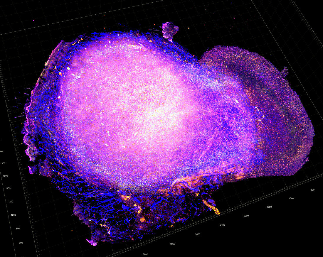
| Breast cancer immune response. 3D transparent tumour tomography image of a breast cancer specimen, showing T cells (yellow, red and blue) attacking the tumour after treatment. This is a HER2-positive breast cancer (human epidermal growth factor receptor 2). The cytotoxic T cells are defined by their cluster of differentiation protein co-receptors: CD3 (yellow), CD8 (red) and CD31 (blue). The tumour had been treated with radiation and a PD-L1 (programmed death-ligand 1) immune checkpoint blockade therapy. Studying how such anti-tumour immune responses can be triggered may lead to better treatments. This image is of a mouse model of breast cancer. | |
| Lizenzart: | Lizenzpflichtig |
| Credit: | Science Photo Library / NATIONAL CANCER INSTITUTE / University of Chicago Comprehensive Cancer Center |
| Bildgröße: | 4704 px × 3744 px |
| Modell-Rechte: | nicht erforderlich |
| Eigentums-Rechte: | nicht erforderlich |
| Restrictions: |
|
Preise für dieses Bild ab 15 €
Universitäten & Organisationen
(Informationsmaterial Digital, Informationsmaterial Print, Lehrmaterial Digital etc.)
ab 15 €
Redaktionell
(Bücher, Bücher: Sach- und Fachliteratur, Digitale Medien (redaktionell) etc.)
ab 30 €
Werbung
(Anzeigen, Aussenwerbung, Digitale Medien, Fernsehwerbung, Karten, Werbemittel, Zeitschriften etc.)
ab 55 €
Handelsprodukte
(bedruckte Textilie, Kalender, Postkarte, Grußkarte, Verpackung etc.)
ab 75 €
Pauschalpreise
Rechtepakete für die unbeschränkte Bildnutzung in Print oder Online
ab 495 €
Keywords
- 3 dimensional,
- 3-dimensional,
- 3D,
- abnormal,
- Behandlungen,
- Brustkrebs,
- cd8,
- Dreidimensional,
- Forschung,
- Gewebe,
- Immunologie,
- immunologisch,
- Immunsystem,
- Kondition,
- Krankheit,
- Krebs,
- krebsartig,
- maligne,
- Malignom,
- Medizin,
- medizinisch,
- Niemand,
- Onkologie,
- Proteine,
- Scanner,
- schwarzer Hintergrund,
- Störung,
- Tumor,
- ungesund,
- Wachstum