Early X-ray arteriogram of injected child,1899
Bildnummer 12100146
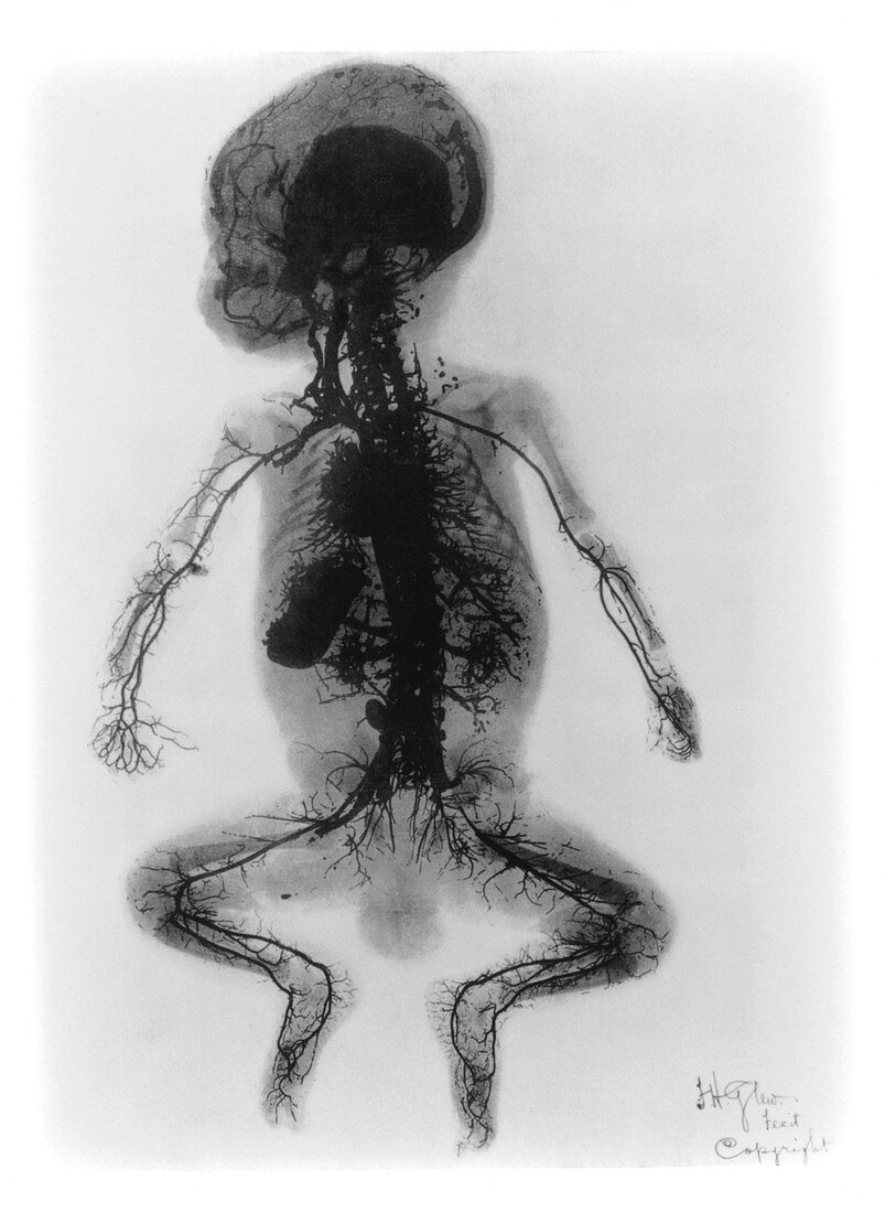
| X-ray arteriogram of a child (1899). Early X-ray showing in fine detail the arteries in a child's body. The X-ray was made in 1899 at St. Thomas' Hospital in London,and published in the Archives of the Roentgen Ray. It was titled: "An injected infant". The cadaver of a young boy was injected,at the femoral artery of the leg,with four pounds of mercury. Under X-ray,arteries of the limbs,abdomen,heart,neck and face are highlighted. This important diagnostic technique was later to become known as an arteriogram or angiogram. It took,however,many years before harmless X-ray opaque substances were developed which could be injected to show arteries in living patients | |
| Lizenzart: | Lizenzpflichtig |
| Credit: | Science Photo Library |
| Bildgröße: | 3607 px × 4942 px |
| Modell-Rechte: | nicht erforderlich |
| Eigentums-Rechte: | nicht erforderlich |
| Restrictions: | - |
Preise für dieses Bild ab 15 €
Universitäten & Organisationen
(Informationsmaterial Digital, Informationsmaterial Print, Lehrmaterial Digital etc.)
ab 15 €
Redaktionell
(Bücher, Bücher: Sach- und Fachliteratur, Digitale Medien (redaktionell) etc.)
ab 30 €
Werbung
(Anzeigen, Aussenwerbung, Digitale Medien, Fernsehwerbung, Karten, Werbemittel, Zeitschriften etc.)
ab 55 €
Handelsprodukte
(bedruckte Textilie, Kalender, Postkarte, Grußkarte, Verpackung etc.)
ab 75 €
Pauschalpreise
Rechtepakete für die unbeschränkte Bildnutzung in Print oder Online
ab 495 €