Pine needle,light micrograph
Bildnummer 12099461
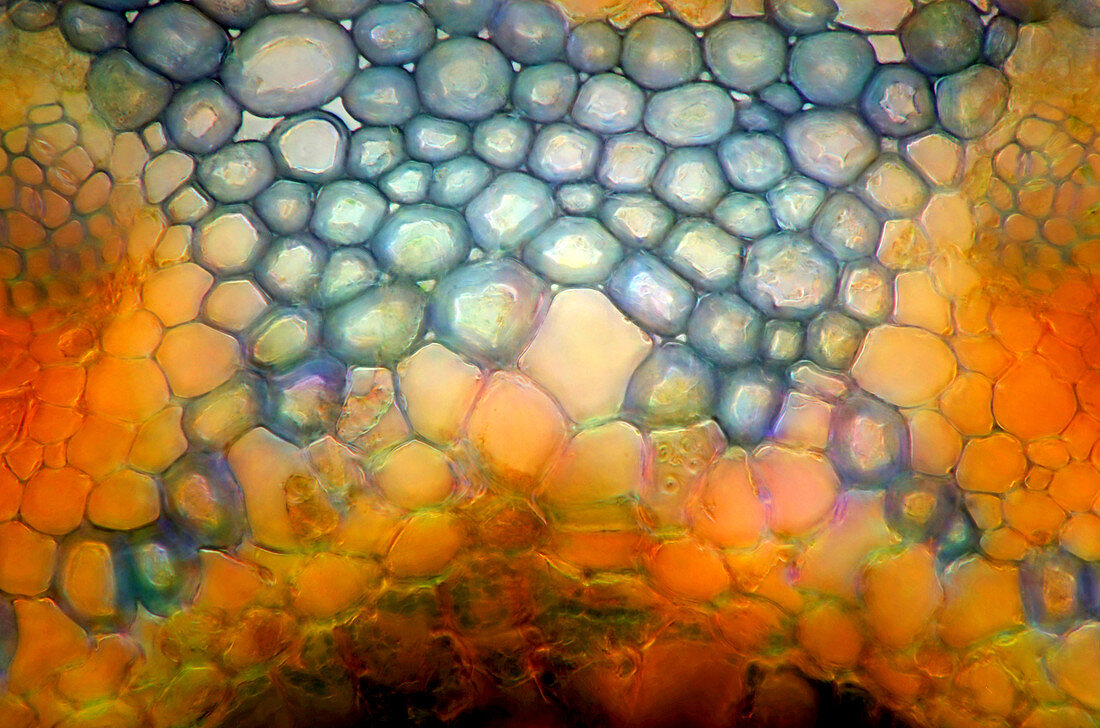
| Pine needle. Darkfield illuminated polarised light micrograph of a cross-section through a pine needle (Pinus sp.). The section shows transfusion tissue within the endodermis in the centre of the needle. Transfusion tissue is composed of parenchyma cells (blue) which fill the space between the vascular bundles (left and right). Magnification: x400,when printed at 10 centimetres wide | |
| Lizenzart: | Lizenzpflichtig |
| Credit: | Science Photo Library / Mis, Marek |
| Bildgröße: | 5143 px × 3406 px |
| Modell-Rechte: | nicht erforderlich |
| Eigentums-Rechte: | nicht erforderlich |
| Restrictions: | - |
Preise für dieses Bild ab 15 €
Universitäten & Organisationen
(Informationsmaterial Digital, Informationsmaterial Print, Lehrmaterial Digital etc.)
ab 15 €
Redaktionell
(Bücher, Bücher: Sach- und Fachliteratur, Digitale Medien (redaktionell) etc.)
ab 30 €
Werbung
(Anzeigen, Aussenwerbung, Digitale Medien, Fernsehwerbung, Karten, Werbemittel, Zeitschriften etc.)
ab 55 €
Handelsprodukte
(bedruckte Textilie, Kalender, Postkarte, Grußkarte, Verpackung etc.)
ab 75 €
Pauschalpreise
Rechtepakete für die unbeschränkte Bildnutzung in Print oder Online
ab 495 €
Keywords
- Anatomie,
- anatomisch,
- Baum,
- Biologie,
- biologisch,
- Blatt,
- Botanik,
- botanisch,
- Bündeln,
- Flora,
- Gefäßband,
- Gewebe,
- gymnospermen,
- Histologie,
- histologisch,
- immergrün,
- Kiefer,
- Licht,
- Lichtmikroskop,
- lichtmikroskopische Aufnahme,
- Mikroskop,
- mikroskopisch,
- Nacktsamer,
- Nadel,
- Nadelbaum,
- Natur,
- Niemand,
- Parenchym,
- Pflanze,
- Pinus,
- polarisiert,
- Querschnitt,
- Transversalschnitt,
- vaskulär,
- zapfentragend,
- Zelle,
- Zellen