Fallopian tube,LM
Bildnummer 12098215
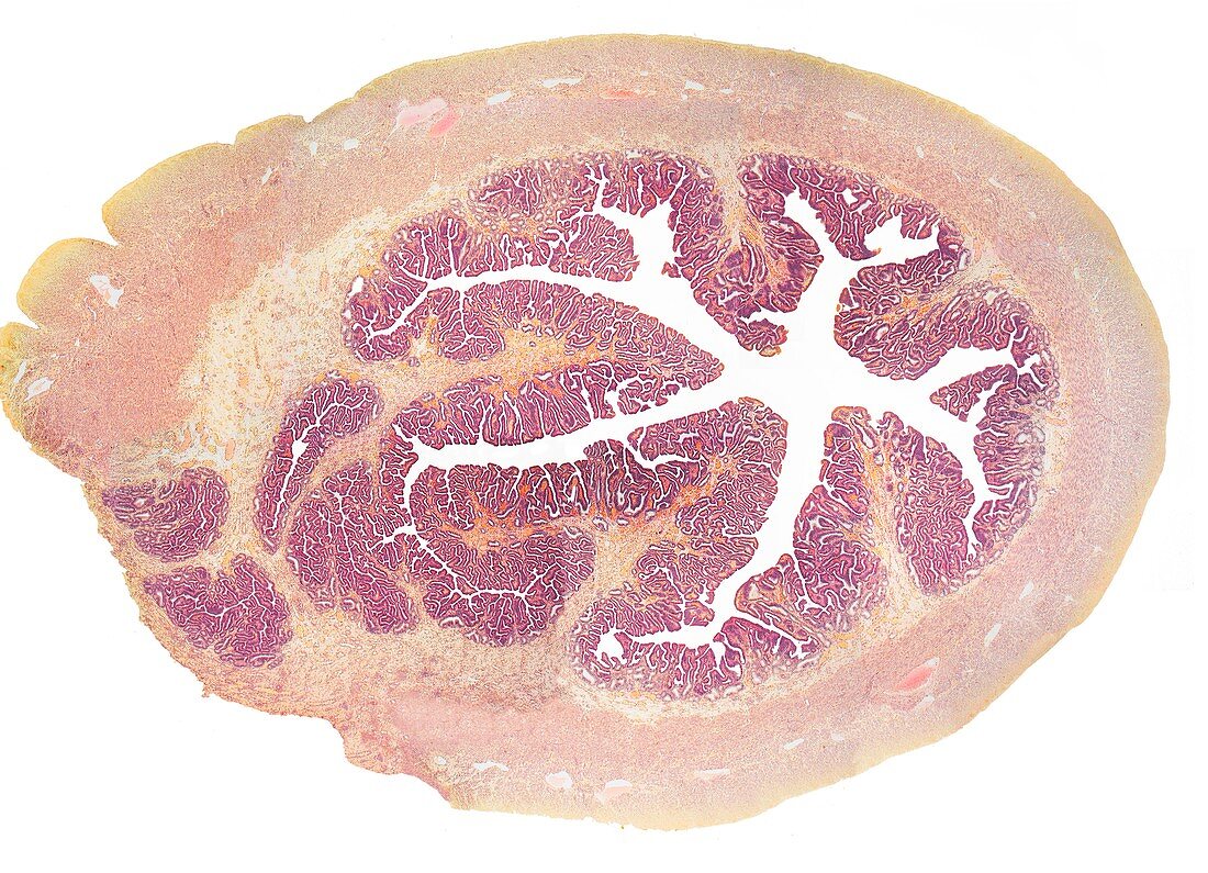
| Fallopian tube. Light micrograph (LM) of a section through a fallopian tube,or oviduct. The fallopian tubes connect the ovaries to the uterus. The lumen is seen centrally,lined with a mucosa (purple) that contains ciliated columnar epithelium and secretory cells. Surrounding this is a vascular layer with capillaries and a layer of smooth muscle (pink).Developing nail. Light micrograph (LM) of longitudinal section through a fetal finger tip to show the developing nail. The large area of pink nail bed epithelium is tipped by the developing nail. Orange connective tissue (dermis) forms the bulk of the image. Pink stratified squamous epithelium of the skin is far left. Magnification: x15 when printed at 10 centimetres wide | |
| Lizenzart: | Lizenzfrei |
| Credit: | Science Photo Library / Gschmeissner, Steve |
| Modell-Rechte: | nicht erforderlich |
| Eigentums-Rechte: | nicht erforderlich |
| Restrictions: | - |
Preise für dieses Bild ab 29 €
Für digitale Nutzung (72 dpi)
ab 29 €
Für Druckauflösung (300 dpi)
ab 300 €