Foetal knee
Bildnummer 12071256
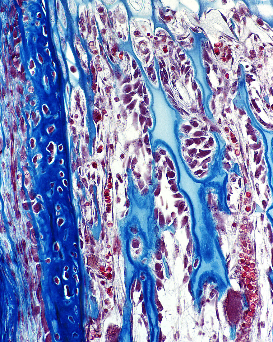
| Light micrograph showing bone and cartilage spicules in the developing long bone (tibia). The spicule at the left side of the micrograph is composed of bone tissue and shows osteocytes embedded in the bone tissue. The lighter appearing spicules consist mostly of calcified cartilage. Numerous osteoblasts are seen in apposition to the cartilage spicules. They are in the process of laying down bone matrix against the calcified cartilage which has a pale blue appearance. An osteoclast is seen at the bottom right of the micrograph. Mallory-Azan stain. Magnification: x350 | |
| Lizenzart: | Lizenzpflichtig |
| Credit: | Science Photo Library / Ross, Michael |
| Bildgröße: | 1500 px × 1880 px |
| Modell-Rechte: | nicht erforderlich |
| Eigentums-Rechte: | nicht erforderlich |
| Restrictions: |
|
Preise für dieses Bild ab 15 €
Universitäten & Organisationen
(Informationsmaterial Digital, Informationsmaterial Print, Lehrmaterial Digital etc.)
ab 15 €
Redaktionell
(Bücher, Bücher: Sach- und Fachliteratur, Digitale Medien (redaktionell) etc.)
ab 30 €
Werbung
(Anzeigen, Aussenwerbung, Digitale Medien, Fernsehwerbung, Karten, Werbemittel, Zeitschriften etc.)
ab 55 €
Handelsprodukte
(bedruckte Textilie, Kalender, Postkarte, Grußkarte, Verpackung etc.)
ab 75 €
Pauschalpreise
Rechtepakete für die unbeschränkte Bildnutzung in Print oder Online
ab 495 €