44-day-old Embryo (Micro-MRI)
Bildnummer 12071241
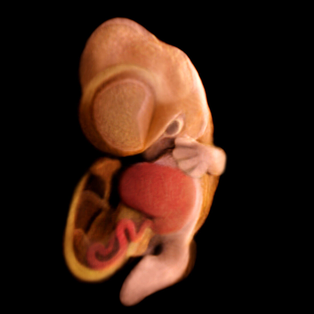
| Computer generated image reconstructed from Micro-MRI,actual size of embryo = 13.0 mm - This head-on view of the human embryo at seven weeks reveals the intricacy in the developing internal organs and structures. Age is calculated from the day of fertilization. Developing after the major organs such as the brain and heart had formed,this image shows the liver (red) in the abdomen region. As well,distinctions from pigmentation can be seen in the left eye. The embryo has now acquired a more human-like appearance,digital rays,precursors to finger formations,can be seen in the hand plate regions,and though the toes have not formed yet,the leg regions have elongated and started to differentiate into thighs and ankles. Image from the book From Conception to Birth: A Life Unfolds | |
| Lizenzart: | Lizenzpflichtig |
| Credit: | Science Photo Library / Anatomical Travelogue |
| Bildgröße: | 3075 px × 3075 px |
| Modell-Rechte: | nicht erforderlich |
| Eigentums-Rechte: | nicht erforderlich |
| Restrictions: |
|
Preise für dieses Bild ab 15 €
Universitäten & Organisationen
(Informationsmaterial Digital, Informationsmaterial Print, Lehrmaterial Digital etc.)
ab 15 €
Redaktionell
(Bücher, Bücher: Sach- und Fachliteratur, Digitale Medien (redaktionell) etc.)
ab 30 €
Werbung
(Anzeigen, Aussenwerbung, Digitale Medien, Fernsehwerbung, Karten, Werbemittel, Zeitschriften etc.)
ab 55 €
Handelsprodukte
(bedruckte Textilie, Kalender, Postkarte, Grußkarte, Verpackung etc.)
ab 75 €
Pauschalpreise
Rechtepakete für die unbeschränkte Bildnutzung in Print oder Online
ab 495 €