Human foetus: stomach,intestine,umbilicus
Bildnummer 12071214
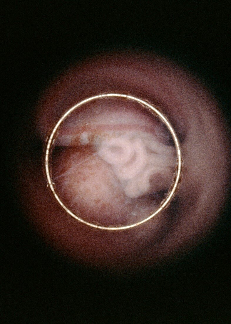
| Endoscopic image of the human foetus in vivo,after 9 weeks,showing: the stomach (bottom,left),the coiled intestines (centre,right),and the umbilicus (top),which carries 2 arteries that remove deoxygenated blood and a vein that provides oxygenated blood for the foetus. The endoscope,a series of lenses and a fibre- optic light guide,is inserted through the cervix and manipulated until a "window" into the amniotic sac is found (ie an area where the wall is thinner) | |
| Lizenzart: | Lizenzpflichtig |
| Credit: | Science Photo Library / Tsiaras, Alexander |
| Bildgröße: | 3621 px × 5070 px |
| Modell-Rechte: | nicht erforderlich |
| Eigentums-Rechte: | nicht erforderlich |
| Restrictions: |
|
Preise für dieses Bild ab 15 €
Universitäten & Organisationen
(Informationsmaterial Digital, Informationsmaterial Print, Lehrmaterial Digital etc.)
ab 15 €
Redaktionell
(Bücher, Bücher: Sach- und Fachliteratur, Digitale Medien (redaktionell) etc.)
ab 30 €
Werbung
(Anzeigen, Aussenwerbung, Digitale Medien, Fernsehwerbung, Karten, Werbemittel, Zeitschriften etc.)
ab 55 €
Handelsprodukte
(bedruckte Textilie, Kalender, Postkarte, Grußkarte, Verpackung etc.)
ab 75 €
Pauschalpreise
Rechtepakete für die unbeschränkte Bildnutzung in Print oder Online
ab 495 €