Middle prophase of mitosis,LM
Bildnummer 12071200
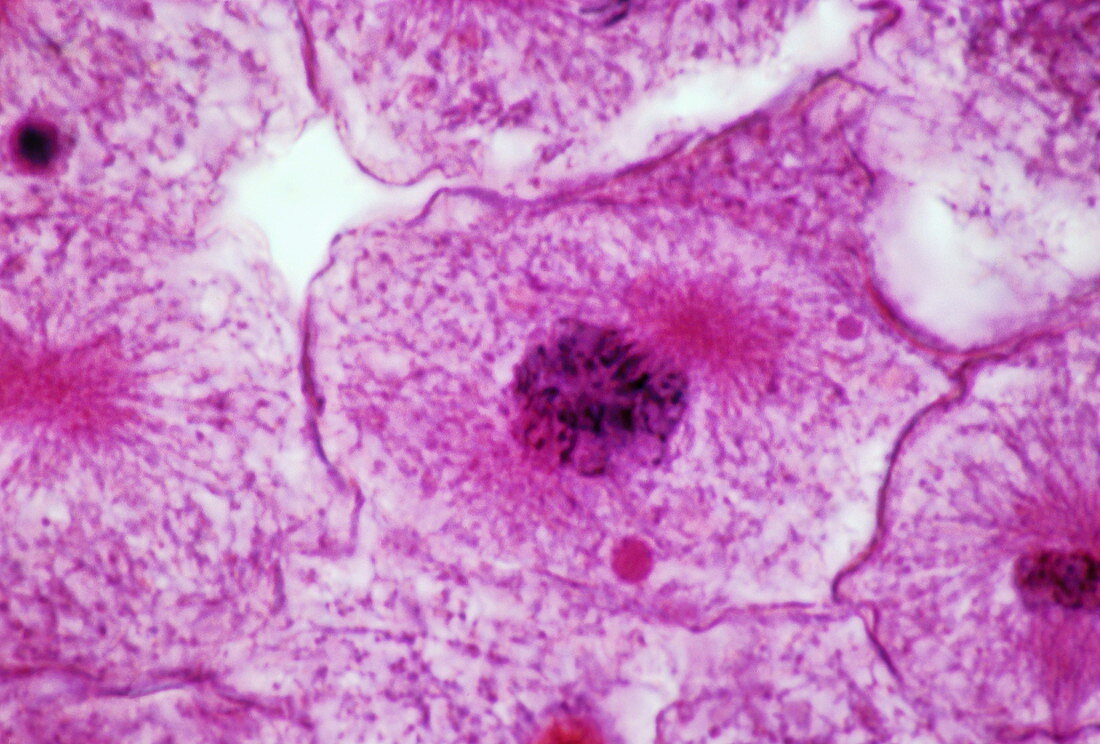
| Middle prophase of mitosis. (Image 2 of 13). Light micrograph of a whitefish cell (Coregonus sp.) undergoing cell division (mitosis). The cell is in the middle prophase (first) stage of mitosis. Individual chromosomes can be seen within the cell as black thread-like structures (centre). During prophase,the chromosomes become progressively shorter and fatter,and a tubular structure known as the spindle forms across the cell (not visible here). The spindle will later help each chromosome to split and divide,forming two 'daughter' nuclei at either end of the cell. For a sequence showing mitosis,see images P673/069-081. Magnification: x900 when printed at 10 centimetres wide | |
| Lizenzart: | Lizenzpflichtig |
| Credit: | Science Photo Library / Abbey, Michael |
| Bildgröße: | 3503 px × 2370 px |
| Modell-Rechte: | nicht erforderlich |
| Eigentums-Rechte: | nicht erforderlich |
| Restrictions: |
|
Preise für dieses Bild ab 15 €
Universitäten & Organisationen
(Informationsmaterial Digital, Informationsmaterial Print, Lehrmaterial Digital etc.)
ab 15 €
Redaktionell
(Bücher, Bücher: Sach- und Fachliteratur, Digitale Medien (redaktionell) etc.)
ab 30 €
Werbung
(Anzeigen, Aussenwerbung, Digitale Medien, Fernsehwerbung, Karten, Werbemittel, Zeitschriften etc.)
ab 55 €
Handelsprodukte
(bedruckte Textilie, Kalender, Postkarte, Grußkarte, Verpackung etc.)
ab 75 €
Pauschalpreise
Rechtepakete für die unbeschränkte Bildnutzung in Print oder Online
ab 495 €