Female reproductive system
Bildnummer 12071049
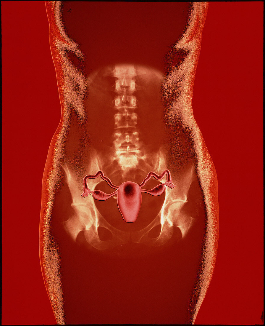
| X-ray & artwork of the female reproductive regions superimposed on the body of a woman. The x-ray shows the pelvic girdle,the ball & socket joints of the hips & the lumbar vertebrae (lower) of the spinal column. The central pear-shaped body is the uterus,or womb. At either side of the uterus,& attached to it by ovarian ligaments,are the two ovaries,where eggs mature. The fallopian tubes which transport eggs from the ovaries to the uterus are headed by flower-shaped structures which guide the eggs into the opening of the tubes. Fertilisation of an egg usually occurs in the tube. Once it reaches the uterus the egg implants in the endometrium,or uterine lining | |
| Lizenzart: | Lizenzpflichtig |
| Credit: | Science Photo Library / Longcore, Bill |
| Bildgröße: | 3457 px × 4242 px |
| Modell-Rechte: | nicht erforderlich |
| Eigentums-Rechte: | nicht erforderlich |
| Restrictions: |
|
Preise für dieses Bild ab 15 €
Universitäten & Organisationen
(Informationsmaterial Digital, Informationsmaterial Print, Lehrmaterial Digital etc.)
ab 15 €
Redaktionell
(Bücher, Bücher: Sach- und Fachliteratur, Digitale Medien (redaktionell) etc.)
ab 30 €
Werbung
(Anzeigen, Aussenwerbung, Digitale Medien, Fernsehwerbung, Karten, Werbemittel, Zeitschriften etc.)
ab 55 €
Handelsprodukte
(bedruckte Textilie, Kalender, Postkarte, Grußkarte, Verpackung etc.)
ab 75 €
Pauschalpreise
Rechtepakete für die unbeschränkte Bildnutzung in Print oder Online
ab 495 €