SEM of an absorptive cell in the small intestine
Bildnummer 12070829
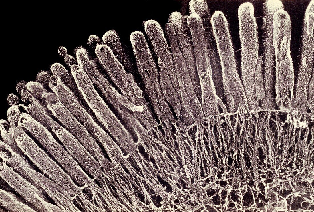
| Scanning electron micrograph (SEM) of an absorptive epithelial cell in the small intestine,showing the parallel arrangement of numerous microvilli that project into the interior lumen of the intestine. The vast surface area provided by the microvilli increases absorption of ingested substances. The deep etching technique used to prepare the specimen also shows the network of microfilaments that radiate from the base,and extend into the core,of each microvillus. The filaments contain the contractile proteins actin & myosin (found in muscle cells),which gives some motility to the microvillus border | |
| Lizenzart: | Lizenzpflichtig |
| Credit: | Science Photo Library / Fawcett / Hirokawa / Heuser |
| Bildgröße: | 5143 px × 3474 px |
| Modell-Rechte: | nicht erforderlich |
| Eigentums-Rechte: | nicht erforderlich |
| Restrictions: |
|
Preise für dieses Bild ab 15 €
Universitäten & Organisationen
(Informationsmaterial Digital, Informationsmaterial Print, Lehrmaterial Digital etc.)
ab 15 €
Redaktionell
(Bücher, Bücher: Sach- und Fachliteratur, Digitale Medien (redaktionell) etc.)
ab 30 €
Werbung
(Anzeigen, Aussenwerbung, Digitale Medien, Fernsehwerbung, Karten, Werbemittel, Zeitschriften etc.)
ab 55 €
Handelsprodukte
(bedruckte Textilie, Kalender, Postkarte, Grußkarte, Verpackung etc.)
ab 75 €
Pauschalpreise
Rechtepakete für die unbeschränkte Bildnutzung in Print oder Online
ab 495 €