Myelination in a newborn
Bildnummer 12070558
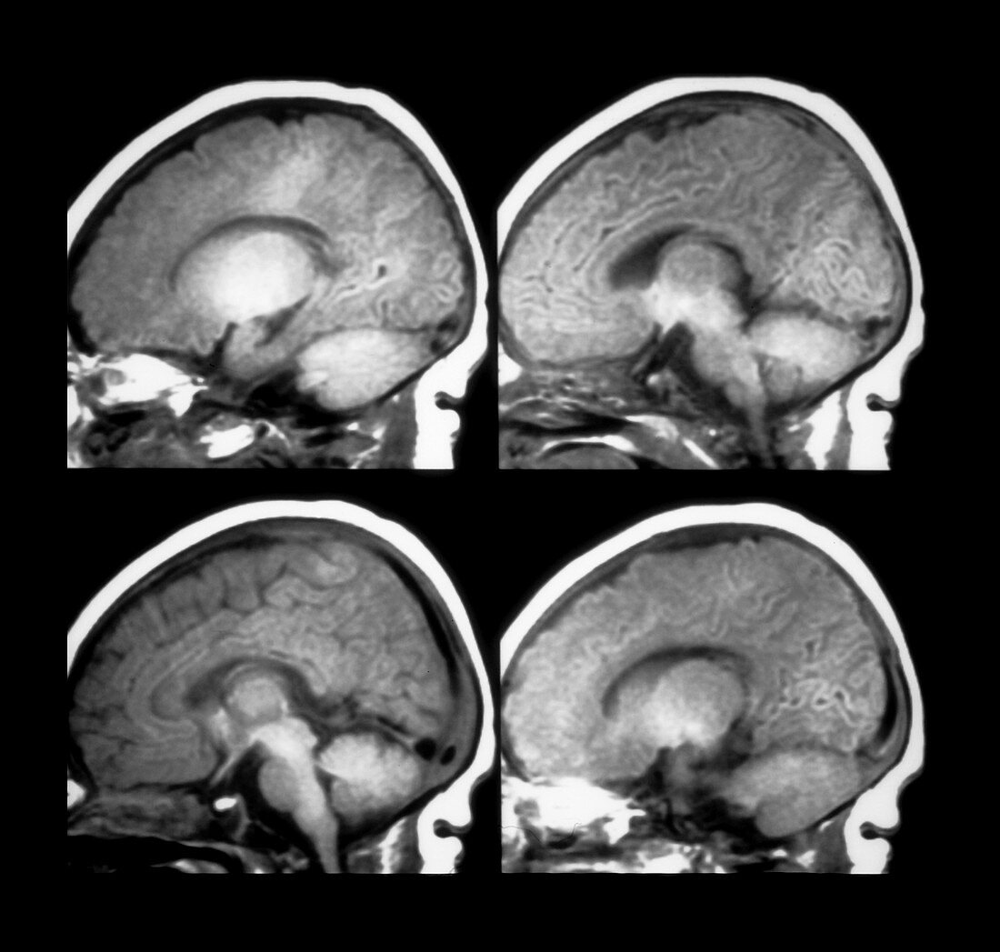
| This composite of 4 axial T1 weighted MRI images of a newborn shows a normal pattern of myelination. The general progression of myelination in infants is from inferior to superior,central to peripheral and posterior to anterior. Immature myelinated regions of the brain have less fat and more water content therefore on T1 weighted images the immature regions of myelination look darker and brighter on T2 weighted images. A relatively normal adult pattern of myelination is acheived by about 18 months of age | |
| Lizenzart: | Lizenzpflichtig |
| Credit: | Science Photo Library / Living Art Enterprises, LLC |
| Bildgröße: | 7545 px × 7183 px |
| Modell-Rechte: | nicht erforderlich |
| Eigentums-Rechte: | nicht erforderlich |
| Restrictions: |
|
Preise für dieses Bild ab 15 €
Universitäten & Organisationen
(Informationsmaterial Digital, Informationsmaterial Print, Lehrmaterial Digital etc.)
ab 15 €
Redaktionell
(Bücher, Bücher: Sach- und Fachliteratur, Digitale Medien (redaktionell) etc.)
ab 30 €
Werbung
(Anzeigen, Aussenwerbung, Digitale Medien, Fernsehwerbung, Karten, Werbemittel, Zeitschriften etc.)
ab 55 €
Handelsprodukte
(bedruckte Textilie, Kalender, Postkarte, Grußkarte, Verpackung etc.)
ab 75 €
Pauschalpreise
Rechtepakete für die unbeschränkte Bildnutzung in Print oder Online
ab 495 €
Keywords
- Anatomie,
- Baby,
- Demyelinisierung,
- Diagnose,
- diagnostische Bildgebung,
- diagnostizieren,
- Gehirn,
- Hirnscan,
- Kopf,
- Magnetresonanzbildgebung,
- Medizin,
- medizinisch,
- medizinische Bildgebung,
- medizinisches Bild,
- Menschen,
- menschlicher Körper,
- MRI,
- Myelinisierung,
- Nerv,
- Nerven,
- Nervensystem,
- Neugeborenes,
- Neuroimaging,
- normal,
- Querschnitt,
- Säugling,
- Verstand,
- Wissenschaft