False-colour MRI image of brain of 80-year-old man
Bildnummer 12070505
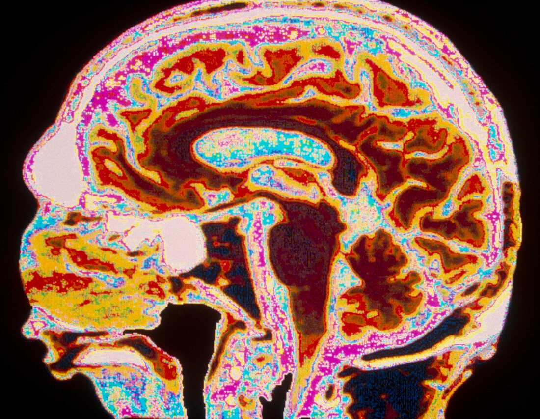
| False-colour magnetic resonance image (MRI) of the head and normal brain of an 80-year-old man. The face is at left: the profile is not complete as this sagittal section is not perfectly central. The brow crest is seen upper left (white). To its right are the cerebral convolutions of the brain (yellow,orange). At centre (brown) is brainstem,which communicates with the spinal cord. Beside the brainstem (right) is the smaller,convoluted cerebellum. MRI imaging is a valuable diagnostic technique which gives good contrast between normal and abnormal tissues (such as tumours). MRI does not use X-rays or other harmful radiation; instead it uses a strong magnetic field and radio waves | |
| Lizenzart: | Lizenzpflichtig |
| Credit: | Science Photo Library / Porett, Thomas |
| Bildgröße: | 5176 px × 4014 px |
| Modell-Rechte: | nicht erforderlich |
| Eigentums-Rechte: | nicht erforderlich |
| Restrictions: |
|
Preise für dieses Bild ab 15 €
Universitäten & Organisationen
(Informationsmaterial Digital, Informationsmaterial Print, Lehrmaterial Digital etc.)
ab 15 €
Redaktionell
(Bücher, Bücher: Sach- und Fachliteratur, Digitale Medien (redaktionell) etc.)
ab 30 €
Werbung
(Anzeigen, Aussenwerbung, Digitale Medien, Fernsehwerbung, Karten, Werbemittel, Zeitschriften etc.)
ab 55 €
Handelsprodukte
(bedruckte Textilie, Kalender, Postkarte, Grußkarte, Verpackung etc.)
ab 75 €
Pauschalpreise
Rechtepakete für die unbeschränkte Bildnutzung in Print oder Online
ab 495 €