Freeze-etched TEM of striated muscle
Bildnummer 12070091
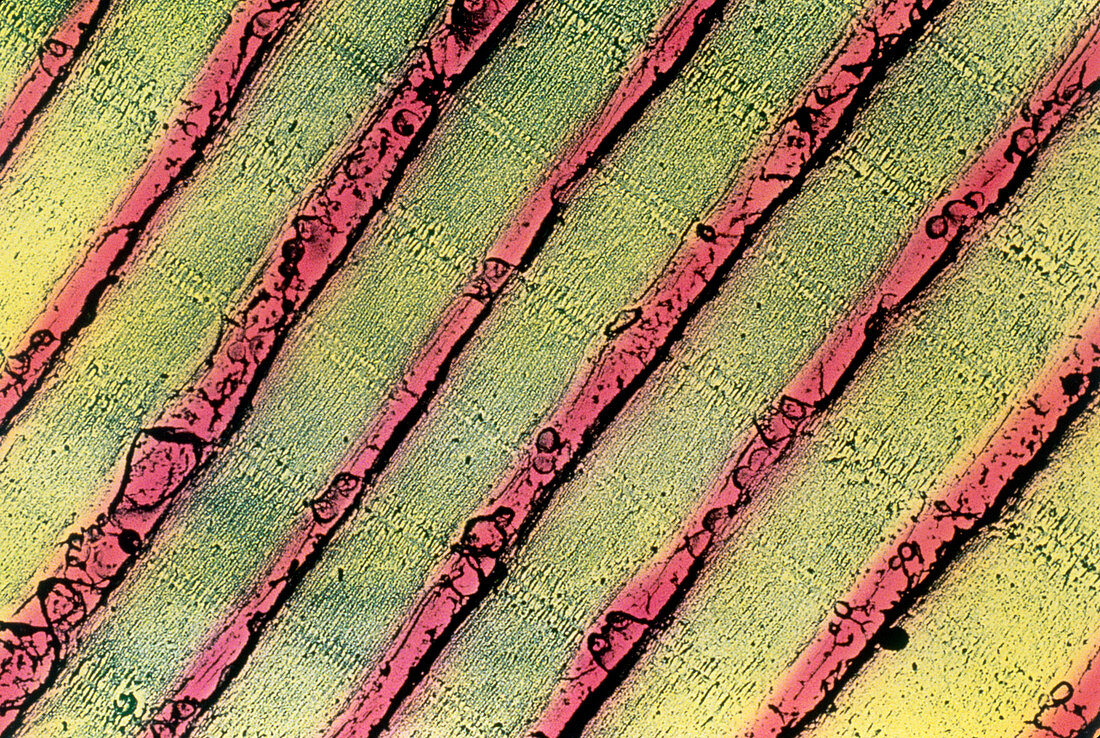
| Muscle cell. Freeze-etched Transmission Electron Micrograph (TEM) of myofibrils (yellow stripes) in a striated (skeletal) muscle cell. Myofibrils con- tain tiny protein filaments that show here as thin black lines running parallel to the direction of the myofibrils. These "myofilaments" consist of two types of protein: actin & myosin. When activated,the actin and myosin filaments slide over each other,overlapping and making the muscle cell contract. Freeze etching involves fracturing deep-frozen cells and then evaporating water from their surface to throw the structure into relief. Magnification unknown | |
| Lizenzart: | Lizenzpflichtig |
| Credit: | Science Photo Library / Franzini Armstrong, Clara |
| Bildgröße: | 5221 px × 3501 px |
| Modell-Rechte: | nicht erforderlich |
| Eigentums-Rechte: | nicht erforderlich |
| Restrictions: |
|
Preise für dieses Bild ab 15 €
Universitäten & Organisationen
(Informationsmaterial Digital, Informationsmaterial Print, Lehrmaterial Digital etc.)
ab 15 €
Redaktionell
(Bücher, Bücher: Sach- und Fachliteratur, Digitale Medien (redaktionell) etc.)
ab 30 €
Werbung
(Anzeigen, Aussenwerbung, Digitale Medien, Fernsehwerbung, Karten, Werbemittel, Zeitschriften etc.)
ab 55 €
Handelsprodukte
(bedruckte Textilie, Kalender, Postkarte, Grußkarte, Verpackung etc.)
ab 75 €
Pauschalpreise
Rechtepakete für die unbeschränkte Bildnutzung in Print oder Online
ab 495 €