X-Ray Showing the Results of an Embolization
Bildnummer 12069252
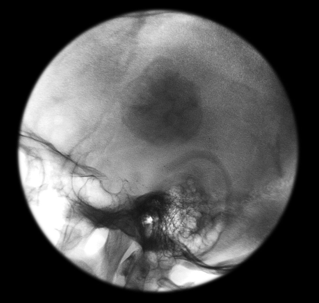
| This lateral ( from the side ) skull x-ray was obtained after the completion of embolization of a convexity meningioma (benign hypervascular tumor). A large amount of small embolic particles which was mixed with xray dye was injected into the blood vessels supplying the intracranial mass (meningioma). After the embolic particles are injected they are trapped in the capillary bed of the tumor which allows them to be seen on a plain x-ray after embolization. Embolization is a way to decrease the blood flow to the tumor to allow the neurosurgeon an easier time when removing it | |
| Lizenzart: | Lizenzpflichtig |
| Credit: | Science Photo Library / Living Art Enterprises, LLC |
| Bildgröße: | 3777 px × 3600 px |
| Modell-Rechte: | nicht erforderlich |
| Eigentums-Rechte: | nicht erforderlich |
| Restrictions: |
|
Preise für dieses Bild ab 15 €
Universitäten & Organisationen
(Informationsmaterial Digital, Informationsmaterial Print, Lehrmaterial Digital etc.)
ab 15 €
Redaktionell
(Bücher, Bücher: Sach- und Fachliteratur, Digitale Medien (redaktionell) etc.)
ab 30 €
Werbung
(Anzeigen, Aussenwerbung, Digitale Medien, Fernsehwerbung, Karten, Werbemittel, Zeitschriften etc.)
ab 55 €
Handelsprodukte
(bedruckte Textilie, Kalender, Postkarte, Grußkarte, Verpackung etc.)
ab 75 €
Pauschalpreise
Rechtepakete für die unbeschränkte Bildnutzung in Print oder Online
ab 495 €