Illustration based on X-ray of an artificial knee
Bildnummer 12069069
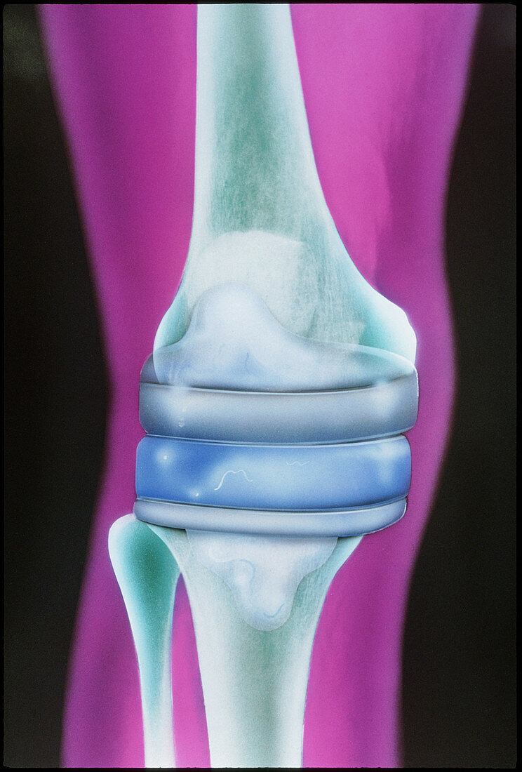
| Prosthetic knee. Illustration based on an X-ray of the artificial knee (at centre) of a 72 year old woman. The two main parts of the replacement knee (grey) are glued into indentations cut out of the femur (thigh bone,upper centre) and tibia (shin- bone,lower centre). A cushion of artificial cartilage or plastic (blue) reduces joint wear. Replacement of the knee joint is most commonly needed to treat osteoarthritis and rheumatoid arthritis | |
| Lizenzart: | Lizenzpflichtig |
| Credit: | Science Photo Library / Bjornberg, Chris |
| Bildgröße: | 3150 px × 4656 px |
| Modell-Rechte: | nicht erforderlich |
| Eigentums-Rechte: | nicht erforderlich |
| Restrictions: |
|
Preise für dieses Bild ab 15 €
Universitäten & Organisationen
(Informationsmaterial Digital, Informationsmaterial Print, Lehrmaterial Digital etc.)
ab 15 €
Redaktionell
(Bücher, Bücher: Sach- und Fachliteratur, Digitale Medien (redaktionell) etc.)
ab 30 €
Werbung
(Anzeigen, Aussenwerbung, Digitale Medien, Fernsehwerbung, Karten, Werbemittel, Zeitschriften etc.)
ab 55 €
Handelsprodukte
(bedruckte Textilie, Kalender, Postkarte, Grußkarte, Verpackung etc.)
ab 75 €
Pauschalpreise
Rechtepakete für die unbeschränkte Bildnutzung in Print oder Online
ab 495 €