Bone fracture
Bildnummer 12068282
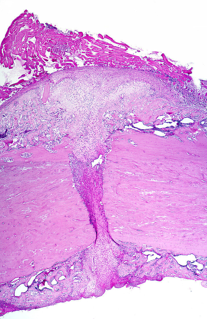
| This micrograph shows the fracture site of a long bone. At the very top of the micrograph is striated muscle. Below this site and at the bottom of the micrograph is the callus of the bone that has been produced in the repair process. The mid-portion of the micrograph shows on either side the original bone at the fracture site with connective tissue and marrow within the fracture. The light staining regions in the callus consist of cartilage which ultimately will be replaced by bone tissue to complete the repair of the fracture. H&E stain. Magnification: x45 | |
| Lizenzart: | Lizenzpflichtig |
| Credit: | Science Photo Library / Ross, Michael |
| Bildgröße: | 1693 px × 2605 px |
| Modell-Rechte: | nicht erforderlich |
| Eigentums-Rechte: | nicht erforderlich |
| Restrictions: |
|
Preise für dieses Bild ab 15 €
Universitäten & Organisationen
(Informationsmaterial Digital, Informationsmaterial Print, Lehrmaterial Digital etc.)
ab 15 €
Redaktionell
(Bücher, Bücher: Sach- und Fachliteratur, Digitale Medien (redaktionell) etc.)
ab 30 €
Werbung
(Anzeigen, Aussenwerbung, Digitale Medien, Fernsehwerbung, Karten, Werbemittel, Zeitschriften etc.)
ab 55 €
Handelsprodukte
(bedruckte Textilie, Kalender, Postkarte, Grußkarte, Verpackung etc.)
ab 75 €
Pauschalpreise
Rechtepakete für die unbeschränkte Bildnutzung in Print oder Online
ab 495 €