Coloured MRI of herniated discs in the neck
Bildnummer 12068237
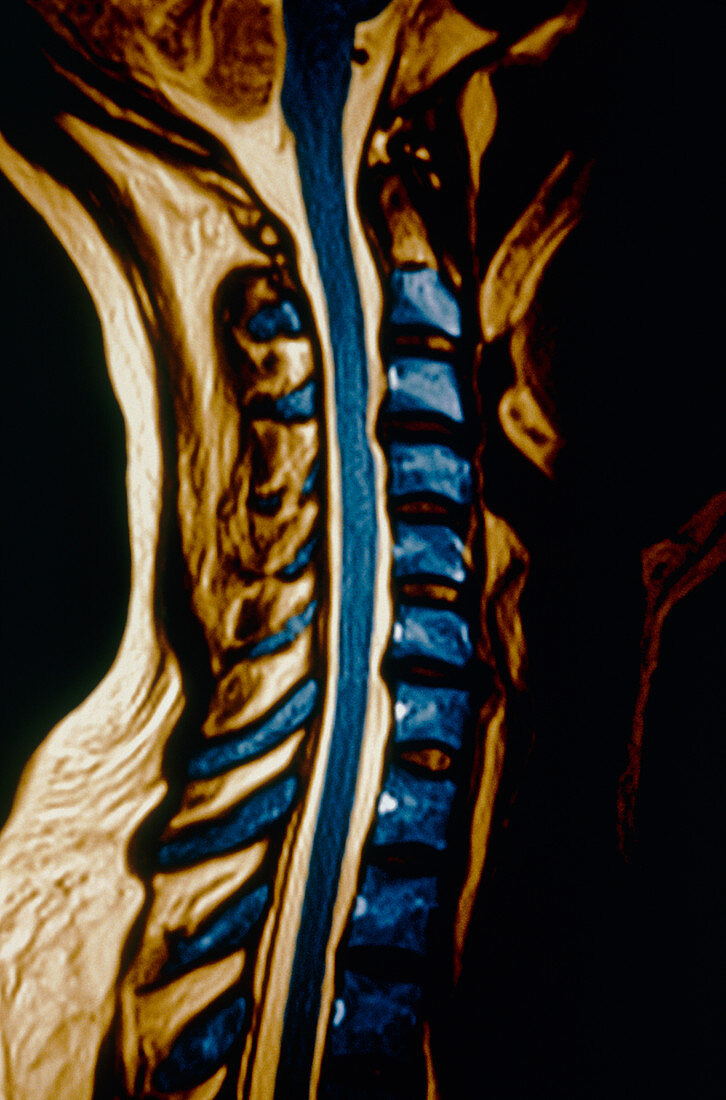
| Herniated discs. Coloured magnetic resonance imag- ing (MRI) scan of a sagittal (side) view through the neck showing herniated (slipped) discs of the spine. The front of the body is at right. The most severely herniated (prolapsed) disc lies between the second (C2) and third (C3) cervical vertebrae (second and third blue blocks seen from the top). The disc has burst and its pulp is indenting the white (pale brown) and grey matter (blue) of the spinal cord (down centre). The disc between C5 & C6 is also herniated. Disc prolapse is often due to discs gradually deteriorating with age. Prolapse causes pain along the affected nerve. While bed rest can help,surgery may be necessary | |
| Lizenzart: | Lizenzpflichtig |
| Credit: | Science Photo Library / Leavines, Susan |
| Bildgröße: | 3376 px × 5112 px |
| Modell-Rechte: | nicht erforderlich |
| Eigentums-Rechte: | nicht erforderlich |
| Restrictions: |
|
Preise für dieses Bild ab 15 €
Universitäten & Organisationen
(Informationsmaterial Digital, Informationsmaterial Print, Lehrmaterial Digital etc.)
ab 15 €
Redaktionell
(Bücher, Bücher: Sach- und Fachliteratur, Digitale Medien (redaktionell) etc.)
ab 30 €
Werbung
(Anzeigen, Aussenwerbung, Digitale Medien, Fernsehwerbung, Karten, Werbemittel, Zeitschriften etc.)
ab 55 €
Handelsprodukte
(bedruckte Textilie, Kalender, Postkarte, Grußkarte, Verpackung etc.)
ab 75 €
Pauschalpreise
Rechtepakete für die unbeschränkte Bildnutzung in Print oder Online
ab 495 €