MRI of Otomastoiditis and Sphenoid Sinusitis
Bildnummer 12068161
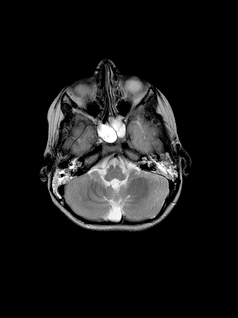
| This T2 weighted axial (cross sectional) MRI image of the head beautifully demonstrates bilateral otomastoiditis and sphenoid sinusitis. There is extensive increased signal intensity (looks white) in the mastoid air cells and middle ear clefts,bilaterally. There is also complete opacification of the sphenoid sinus,which also demonstrates prominent abnormal increased signal intensity (looks white) | |
| Lizenzart: | Lizenzpflichtig |
| Credit: | Science Photo Library / Living Art Enterprises, LLC |
| Bildgröße: | 2700 px × 3600 px |
| Modell-Rechte: | nicht erforderlich |
| Eigentums-Rechte: | nicht erforderlich |
| Restrictions: |
|
Preise für dieses Bild ab 15 €
Universitäten & Organisationen
(Informationsmaterial Digital, Informationsmaterial Print, Lehrmaterial Digital etc.)
ab 15 €
Redaktionell
(Bücher, Bücher: Sach- und Fachliteratur, Digitale Medien (redaktionell) etc.)
ab 30 €
Werbung
(Anzeigen, Aussenwerbung, Digitale Medien, Fernsehwerbung, Karten, Werbemittel, Zeitschriften etc.)
ab 55 €
Handelsprodukte
(bedruckte Textilie, Kalender, Postkarte, Grußkarte, Verpackung etc.)
ab 75 €
Pauschalpreise
Rechtepakete für die unbeschränkte Bildnutzung in Print oder Online
ab 495 €