MRI of Glial tumour-
Bildnummer 12067476
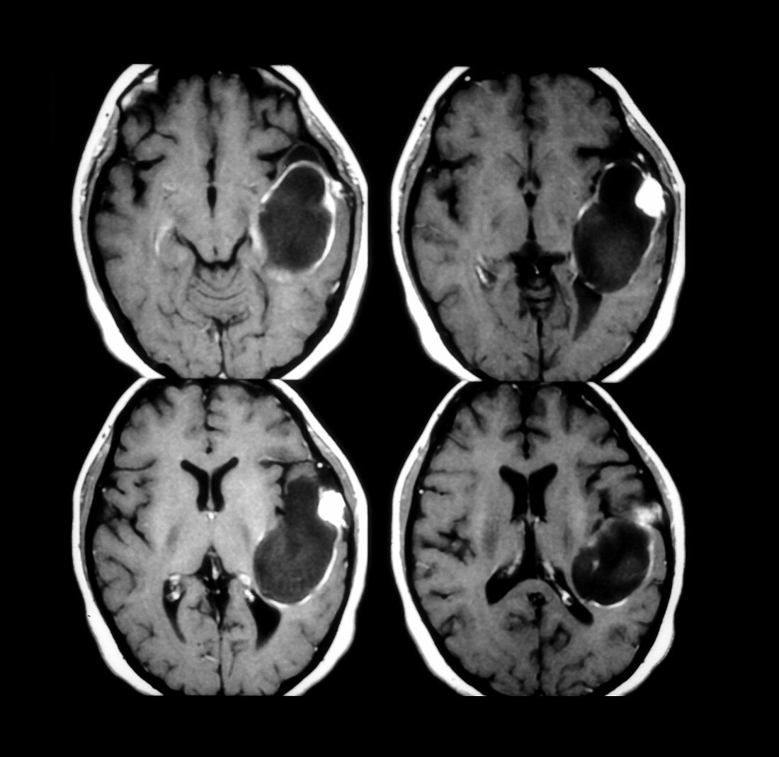
| This composite of 4 axial (cross sectional) contrast enhanced T1 weighted MRI images of a 13 year old male with seizures shows the typical appearance of this unusual type of glial (astrocytic) tumour- which often is located in the temporal lobes and often consists of a cyst with a mural nodue as this case demonstrates. This tumour- is most often found in children and young adults,is often associated with seizures and may involve the leptomeninges | |
| Lizenzart: | Lizenzpflichtig |
| Credit: | Science Photo Library / Living Art Enterprises, LLC |
| Bildgröße: | 4944 px × 4801 px |
| Modell-Rechte: | nicht erforderlich |
| Eigentums-Rechte: | nicht erforderlich |
| Restrictions: |
|
Preise für dieses Bild ab 15 €
Universitäten & Organisationen
(Informationsmaterial Digital, Informationsmaterial Print, Lehrmaterial Digital etc.)
ab 15 €
Redaktionell
(Bücher, Bücher: Sach- und Fachliteratur, Digitale Medien (redaktionell) etc.)
ab 30 €
Werbung
(Anzeigen, Aussenwerbung, Digitale Medien, Fernsehwerbung, Karten, Werbemittel, Zeitschriften etc.)
ab 55 €
Handelsprodukte
(bedruckte Textilie, Kalender, Postkarte, Grußkarte, Verpackung etc.)
ab 75 €
Pauschalpreise
Rechtepakete für die unbeschränkte Bildnutzung in Print oder Online
ab 495 €
Keywords
- Bild,
- Diagnose,
- diagnostische Bildgebung,
- diagnostizieren,
- Gehirn,
- Gesundheitswesen,
- Hirntumor,
- Kinder,
- Kondition,
- Krankheit,
- Krebs,
- krebsartig,
- maligne,
- Malignom,
- Medizin,
- medizinisch,
- medizinische Bildgebung,
- medizinischer Scan,
- medizinisches Bild,
- Neuroimaging,
- Störung,
- Temporallappen,
- Wissenschaft