Stomach cilia,TEM
Bildnummer 12050111
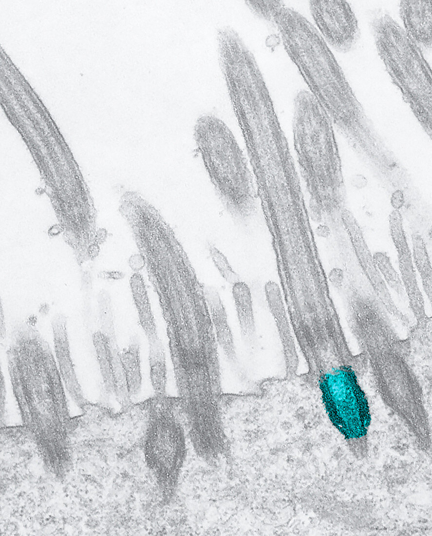
| Stomach cilia. Coloured transmission electron micrograph (TEM) of a section through the brush border lining in the stomach wall of a house mouse (Mus musculus),showing the cilia (hair-like) that cover its surface. Here,the basal body of one of the cilia is shown (purple). The basal body is the site of growth for the microtubules that make up the axoneme,which provides support and facilitates movement in each cilium | |
| Lizenzart: | Lizenzpflichtig |
| Credit: | Science Photo Library / Miller, Marian |
| Bildgröße: | 2653 px × 3291 px |
| Modell-Rechte: | nicht erforderlich |
| Eigentums-Rechte: | nicht erforderlich |
| Restrictions: | - |
Preise für dieses Bild ab 15 €
Universitäten & Organisationen
(Informationsmaterial Digital, Informationsmaterial Print, Lehrmaterial Digital etc.)
ab 15 €
Redaktionell
(Bücher, Bücher: Sach- und Fachliteratur, Digitale Medien (redaktionell) etc.)
ab 30 €
Werbung
(Anzeigen, Aussenwerbung, Digitale Medien, Fernsehwerbung, Karten, Werbemittel, Zeitschriften etc.)
ab 55 €
Handelsprodukte
(bedruckte Textilie, Kalender, Postkarte, Grußkarte, Verpackung etc.)
ab 75 €
Pauschalpreise
Rechtepakete für die unbeschränkte Bildnutzung in Print oder Online
ab 495 €
Keywords
- Anatomie,
- anatomisch,
- Bauch,
- Biologie,
- biologisch,
- Bürstensaum,
- Darm,
- Darm-,
- Epithel,
- epithelial,
- Farbig,
- Fauna,
- Forschung,
- Gedärme,
- gefärbt,
- GI tract,
- Histologie,
- histologisch,
- Innere,
- Magen-,
- Magen-Darm-System,
- Natur,
- Niemand,
- Oberfläche,
- Organelle,
- Organellen,
- Schleimhaut,
- Sektion,
- sektioniert,
- tem,
- Tiere,
- tierisches Gewebe,
- Trakt,
- transmissionselektronenmikroskopische Aufnahme,
- Verdauungs-,
- Weiblich,
- Wimpern,
- Zelle,
- Zellen,
- Zoologie,
- zoologisch,
- Zytologie,
- Zytologisch,
- Zytoskelett