Membrane Ultrastructure in Nerve Cells
Bildnummer 12047436
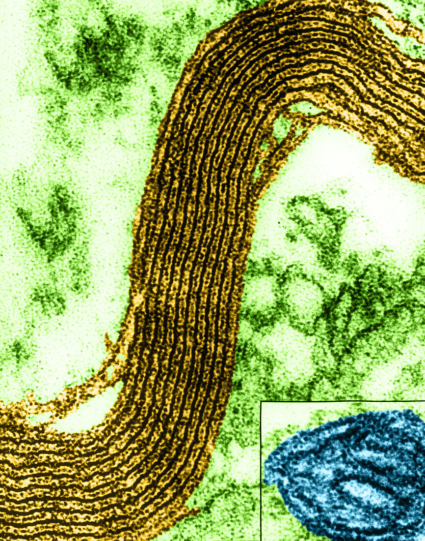
| Colour enhanced high resolution electron micrograph of myelin sheath segment from transverse section of frog sciatic nerve. The main image shows a concentric array of dense and intermediate layers. Particulate sub-unit structures are regularly found within the plane of the layers in these well-preserved osmium fixed,low-temperature preparations. Magnification 480,000x at 9x11 inches. Image B (lower right corner) is a cristae. Magnification 600,000x | |
| Lizenzart: | Lizenzpflichtig |
| Credit: | Science Photo Library / Omikron / Fernandez-Moran |
| Bildgröße: | 3363 px × 4272 px |
| Modell-Rechte: | nicht erforderlich |
| Eigentums-Rechte: | nicht erforderlich |
| Restrictions: |
|
Preise für dieses Bild ab 15 €
Universitäten & Organisationen
(Informationsmaterial Digital, Informationsmaterial Print, Lehrmaterial Digital etc.)
ab 15 €
Redaktionell
(Bücher, Bücher: Sach- und Fachliteratur, Digitale Medien (redaktionell) etc.)
ab 30 €
Werbung
(Anzeigen, Aussenwerbung, Digitale Medien, Fernsehwerbung, Karten, Werbemittel, Zeitschriften etc.)
ab 55 €
Handelsprodukte
(bedruckte Textilie, Kalender, Postkarte, Grußkarte, Verpackung etc.)
ab 75 €
Pauschalpreise
Rechtepakete für die unbeschränkte Bildnutzung in Print oder Online
ab 495 €