Pinocytosis,Capillary Endothelium,TEM
Bildnummer 12047096
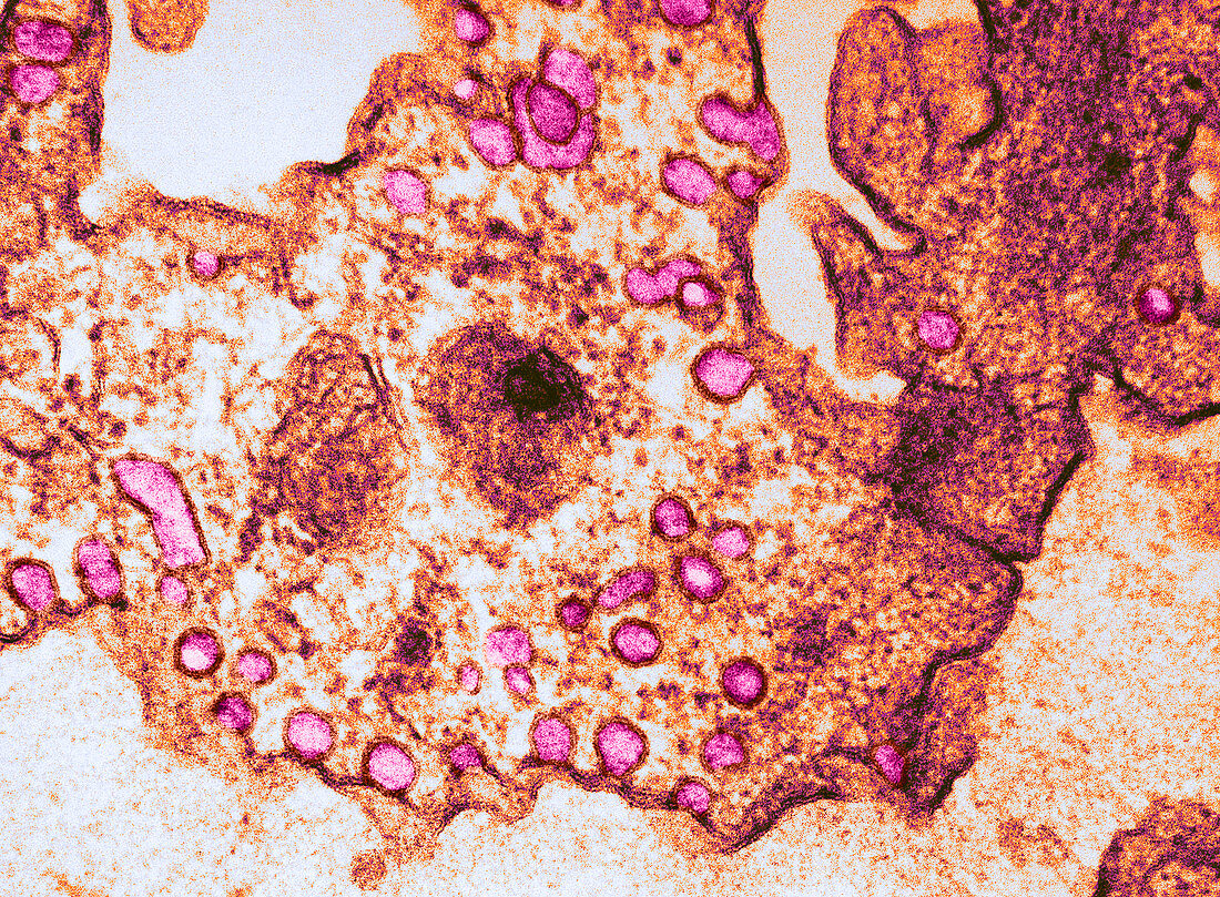
| Colour enhanced transmission electron micrograph of capillary endothelium. Pinocytosing vesicles on the luminal surface (fuchsia) participate in the uptake of material to be transported. Within the cytoplasm of the cell can be seen vesicles in transport while on the plasma membrane facing the lower portion of the picture the material is being released via the process of exocytosis. Magnification 157,000x | |
| Lizenzart: | Lizenzpflichtig |
| Credit: | Science Photo Library / Gennaro, Joseph F. |
| Bildgröße: | 3714 px × 2730 px |
| Modell-Rechte: | nicht erforderlich |
| Eigentums-Rechte: | nicht erforderlich |
| Restrictions: |
|
Preise für dieses Bild ab 15 €
Universitäten & Organisationen
(Informationsmaterial Digital, Informationsmaterial Print, Lehrmaterial Digital etc.)
ab 15 €
Redaktionell
(Bücher, Bücher: Sach- und Fachliteratur, Digitale Medien (redaktionell) etc.)
ab 30 €
Werbung
(Anzeigen, Aussenwerbung, Digitale Medien, Fernsehwerbung, Karten, Werbemittel, Zeitschriften etc.)
ab 55 €
Handelsprodukte
(bedruckte Textilie, Kalender, Postkarte, Grußkarte, Verpackung etc.)
ab 75 €
Pauschalpreise
Rechtepakete für die unbeschränkte Bildnutzung in Print oder Online
ab 495 €
Keywords
- Biologie,
- eingefärbt,
- elektronenmikroskopische Aufnahme,
- em,
- Eukaryot,
- Exozytose,
- Histologie,
- Mikrofotografie,
- Mikrographie,
- Mikroskopie,
- mikroskopisch,
- Niemand,
- Organelle,
- Physiologie,
- Pinozytose,
- tem,
- Transmissionselektronen,
- verbessert,
- Vesikel,
- Zelle,
- Zellstruktur,
- zellular,
- Zytologie,
- Zytoplasma