Giardia sp. Protozoan (TEM)
Bildnummer 12045882
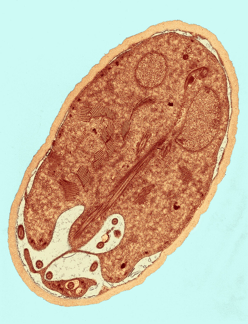
| Colour-enhanced Transmission Electron Micrograph (TEM) showing the cyst-stage of a Giardia sp. protozoan. The outer cyst wall is composed of filamentous and membranous portions,and is separated from the cytoplasm of the trophozoites contained within by the peritrophic space. This cyst wall is approximately 0.25 microns thick | |
| Lizenzart: | Lizenzpflichtig |
| Credit: | Science Photo Library / Science Source |
| Bildgröße: | 1680 px × 2195 px |
| Modell-Rechte: | nicht erforderlich |
| Eigentums-Rechte: | nicht erforderlich |
| Restrictions: |
|
Preise für dieses Bild ab 15 €
Universitäten & Organisationen
(Informationsmaterial Digital, Informationsmaterial Print, Lehrmaterial Digital etc.)
ab 15 €
Redaktionell
(Bücher, Bücher: Sach- und Fachliteratur, Digitale Medien (redaktionell) etc.)
ab 30 €
Werbung
(Anzeigen, Aussenwerbung, Digitale Medien, Fernsehwerbung, Karten, Werbemittel, Zeitschriften etc.)
ab 55 €
Handelsprodukte
(bedruckte Textilie, Kalender, Postkarte, Grußkarte, Verpackung etc.)
ab 75 €
Pauschalpreise
Rechtepakete für die unbeschränkte Bildnutzung in Print oder Online
ab 495 €
Keywords
- abnormal,
- ansteckend,
- Bildgebung,
- eingefärbt,
- elektronenmikroskopische Aufnahme,
- em,
- Erweiterung,
- Farbverbesserung,
- Fehlfarbe,
- Giardiasis,
- Histopathologie,
- Infektion,
- Kolorieren,
- Kondition,
- Krankheit,
- medizinisch,
- Mikrofotografie,
- Mikrographie,
- Niemand,
- Organismus,
- parasitär,
- Pathologie,
- Protozoen,
- Protozoon,
- tem,
- transmissionselektronenmikroskopische Aufnahme,
- ungesund,
- verbessert,
- Wissenschaft,
- Zyste