Micropinocytosis,TEM
Bildnummer 12045636
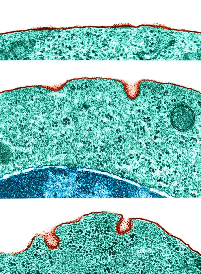
| Colour enhanced transmission electron micrographs of portions of the surface of polychromatophilic erythroblasts from guinea pig bone marrow. This series of images illustrates the intermediate stages in the formation of coated micropinocytotic vesicles. We see small thickened areas of membrane acquire an inner coat and a fuzzy external layer to which particles of ferritin adhere | |
| Lizenzart: | Lizenzpflichtig |
| Credit: | Science Photo Library / Fawcett, Don W. |
| Bildgröße: | 3460 px × 4718 px |
| Modell-Rechte: | nicht erforderlich |
| Eigentums-Rechte: | nicht erforderlich |
| Restrictions: |
|
Preise für dieses Bild ab 15 €
Universitäten & Organisationen
(Informationsmaterial Digital, Informationsmaterial Print, Lehrmaterial Digital etc.)
ab 15 €
Redaktionell
(Bücher, Bücher: Sach- und Fachliteratur, Digitale Medien (redaktionell) etc.)
ab 30 €
Werbung
(Anzeigen, Aussenwerbung, Digitale Medien, Fernsehwerbung, Karten, Werbemittel, Zeitschriften etc.)
ab 55 €
Handelsprodukte
(bedruckte Textilie, Kalender, Postkarte, Grußkarte, Verpackung etc.)
ab 75 €
Pauschalpreise
Rechtepakete für die unbeschränkte Bildnutzung in Print oder Online
ab 495 €