Gap Junctions,TEM
Bildnummer 12045522
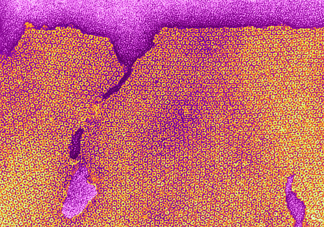
| Colour enhanced transmission electron micrograph of freeze-fractured gap junctions. Gap junctions are isolated from mouse liver cells and examined by negative staining using phosphotungstate or uranyl formate as the contrast medium. At high magnification,the 8 to 9 nm particles or connexons have central 1.5 to 2nm electron-dense region which is believed to represent the polar channel through which cell to cell communication takes place | |
| Lizenzart: | Lizenzpflichtig |
| Credit: | Science Photo Library / Fawcett, Don W. |
| Bildgröße: | 5158 px × 3618 px |
| Modell-Rechte: | nicht erforderlich |
| Eigentums-Rechte: | nicht erforderlich |
| Restrictions: |
|
Preise für dieses Bild ab 15 €
Universitäten & Organisationen
(Informationsmaterial Digital, Informationsmaterial Print, Lehrmaterial Digital etc.)
ab 15 €
Redaktionell
(Bücher, Bücher: Sach- und Fachliteratur, Digitale Medien (redaktionell) etc.)
ab 30 €
Werbung
(Anzeigen, Aussenwerbung, Digitale Medien, Fernsehwerbung, Karten, Werbemittel, Zeitschriften etc.)
ab 55 €
Handelsprodukte
(bedruckte Textilie, Kalender, Postkarte, Grußkarte, Verpackung etc.)
ab 75 €
Pauschalpreise
Rechtepakete für die unbeschränkte Bildnutzung in Print oder Online
ab 495 €