Epidural Space
Bildnummer 12042518
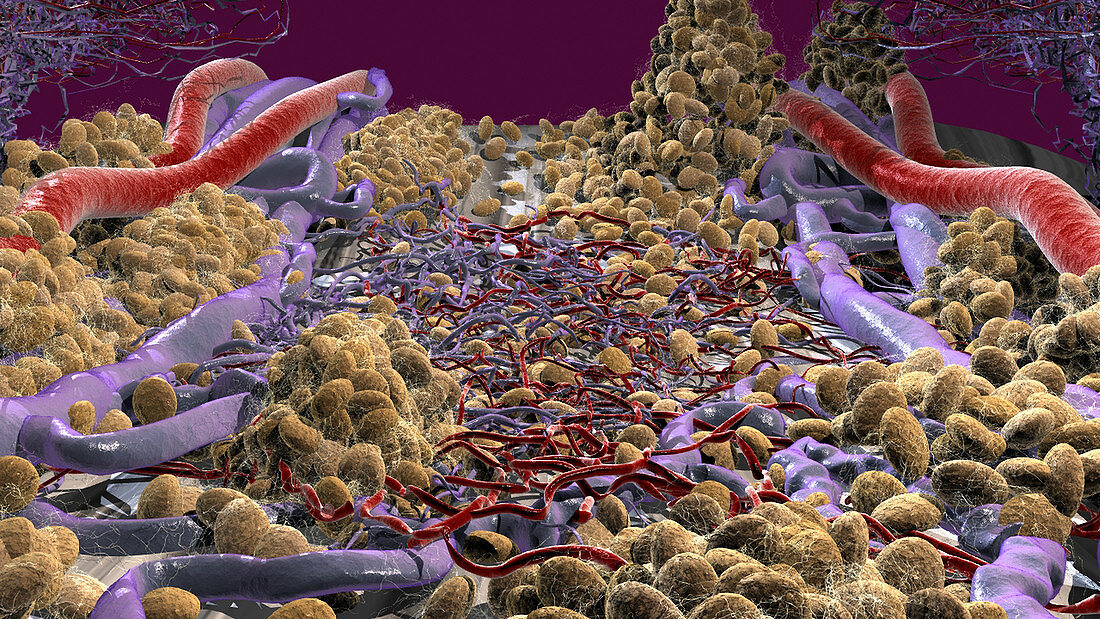
| This microscopic view of the epidural space demonstrates the dense venous plexus and capillaries present throughout this space. Fat cells (yellow) are seen scattered throughout the space connected by connective tissue. The posterior longitudinal ligament is barely visible in the background under fat cells at the top of the image | |
| Lizenzart: | Lizenzpflichtig |
| Credit: | Science Photo Library / Orthoclick |
| Bildgröße: | 4552 px × 2561 px |
| Modell-Rechte: | nicht erforderlich |
| Eigentums-Rechte: | nicht erforderlich |
| Restrictions: |
|
Preise für dieses Bild ab 15 €
Universitäten & Organisationen
(Informationsmaterial Digital, Informationsmaterial Print, Lehrmaterial Digital etc.)
ab 15 €
Redaktionell
(Bücher, Bücher: Sach- und Fachliteratur, Digitale Medien (redaktionell) etc.)
ab 30 €
Werbung
(Anzeigen, Aussenwerbung, Digitale Medien, Fernsehwerbung, Karten, Werbemittel, Zeitschriften etc.)
ab 55 €
Handelsprodukte
(bedruckte Textilie, Kalender, Postkarte, Grußkarte, Verpackung etc.)
ab 75 €
Pauschalpreise
Rechtepakete für die unbeschränkte Bildnutzung in Print oder Online
ab 495 €