Structure of Taste Bud,Illustration
Bildnummer 12041227
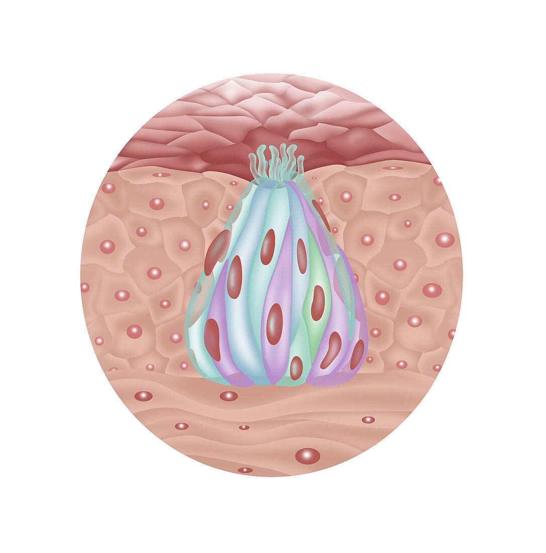
| Illustration of the structure of a taste bud. From top to bottom: Taste pore (dark pink area),gustatory hair (light green follicles),gustatory receptor cell (green & blue panels in pod),basal cell (reddish spots),stratified squamous epithelium (outer light pink area),supporting cell (purple areas),connective tissue (light pink area at bottom),sensory neurons | |
| Lizenzart: | Lizenzpflichtig |
| Credit: | Science Photo Library / Shockey, Gwen |
| Bildgröße: | 1920 px × 1944 px |
| Modell-Rechte: | nicht erforderlich |
| Eigentums-Rechte: | nicht erforderlich |
| Restrictions: |
|
Preise für dieses Bild ab 15 €
Universitäten & Organisationen
(Informationsmaterial Digital, Informationsmaterial Print, Lehrmaterial Digital etc.)
ab 15 €
Redaktionell
(Bücher, Bücher: Sach- und Fachliteratur, Digitale Medien (redaktionell) etc.)
ab 30 €
Werbung
(Anzeigen, Aussenwerbung, Digitale Medien, Fernsehwerbung, Karten, Werbemittel, Zeitschriften etc.)
ab 55 €
Handelsprodukte
(bedruckte Textilie, Kalender, Postkarte, Grußkarte, Verpackung etc.)
ab 75 €
Pauschalpreise
Rechtepakete für die unbeschränkte Bildnutzung in Print oder Online
ab 495 €