MRI of Normal and AVM Brains
Bildnummer 12037116
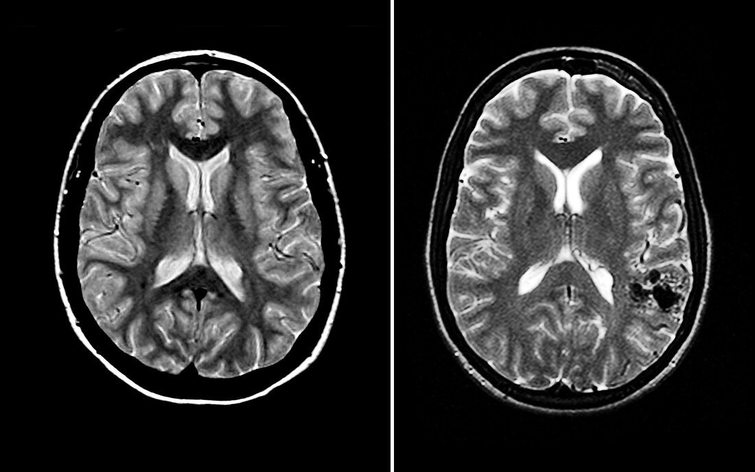
| On the left is a normal cross-sectional MRI image of the brain through both cerebral hemispheres. At this level you see two large cavities filled with dark material. These are the lateral ventricles which are filled with cerebral spinal fluid (CSF). There are two main types of brain tissue,grey matter (which contains the neuronal cell bodies and is the darker of the brain tissue shown) and white matter (which is composed of axonal fibres). On the right is a cross-sectional MRI image of the brain showing the typical appearance of a high-flow arterial venous malformation (AVM). This is depicted as the rounded area of black signal. This is located in the temporal-parietal region. This is a type of congenital brain vascular malformation that can result in serious disability or death. These can rupture (burst) and lead to bleeding inside the head | |
| Lizenzart: | Lizenzpflichtig |
| Credit: | Science Photo Library / Wilson, Jessica |
| Bildgröße: | 5344 px × 3344 px |
| Modell-Rechte: | nicht erforderlich |
| Eigentums-Rechte: | nicht erforderlich |
| Restrictions: |
|
Preise für dieses Bild ab 15 €
Universitäten & Organisationen
(Informationsmaterial Digital, Informationsmaterial Print, Lehrmaterial Digital etc.)
ab 15 €
Redaktionell
(Bücher, Bücher: Sach- und Fachliteratur, Digitale Medien (redaktionell) etc.)
ab 30 €
Werbung
(Anzeigen, Aussenwerbung, Digitale Medien, Fernsehwerbung, Karten, Werbemittel, Zeitschriften etc.)
ab 55 €
Handelsprodukte
(bedruckte Textilie, Kalender, Postkarte, Grußkarte, Verpackung etc.)
ab 75 €
Pauschalpreise
Rechtepakete für die unbeschränkte Bildnutzung in Print oder Online
ab 495 €