MRI of Normal Brain and Acute MS
Bildnummer 12037091
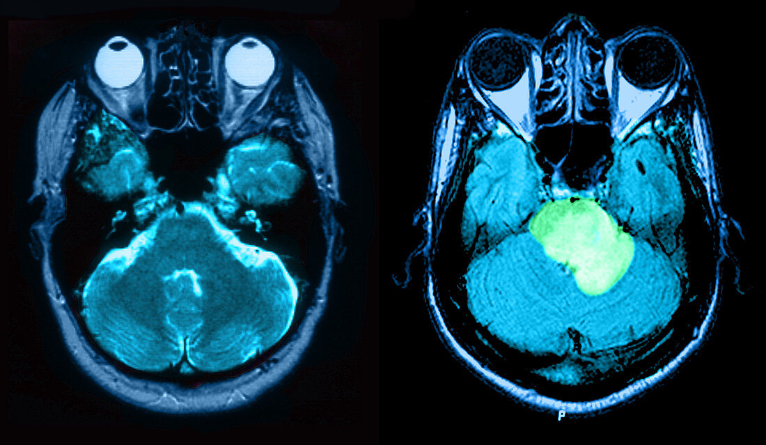
| MRI of normal brain (left) and one with acute multiple sclerosis (right). On the left is an MRI scan,T2 weighted,axial view through the brain of a 54 year old female. On the right is an axial MRI image of the brain through the level of the brain stem,revealing extensive edema and enlargement of the pons (in green) extending into the left (on your right) middle cerebellar peduncle. There is prominent ring enhancement on post contrast images. This represents a form of acute MS that can stimulate a tumour; therefore,it is referred to as tumefactive | |
| Lizenzart: | Lizenzpflichtig |
| Credit: | Science Photo Library / Wilson, Jessica |
| Bildgröße: | 5000 px × 2905 px |
| Modell-Rechte: | nicht erforderlich |
| Eigentums-Rechte: | nicht erforderlich |
| Restrictions: |
|
Preise für dieses Bild ab 15 €
Universitäten & Organisationen
(Informationsmaterial Digital, Informationsmaterial Print, Lehrmaterial Digital etc.)
ab 15 €
Redaktionell
(Bücher, Bücher: Sach- und Fachliteratur, Digitale Medien (redaktionell) etc.)
ab 30 €
Werbung
(Anzeigen, Aussenwerbung, Digitale Medien, Fernsehwerbung, Karten, Werbemittel, Zeitschriften etc.)
ab 55 €
Handelsprodukte
(bedruckte Textilie, Kalender, Postkarte, Grußkarte, Verpackung etc.)
ab 75 €
Pauschalpreise
Rechtepakete für die unbeschränkte Bildnutzung in Print oder Online
ab 495 €